. 2013 May 31;288(22):16196-205.
doi: 10.1074/jbc.M112.435461. Epub 2013 Apr 18.

Innate immunity in pluripotent human cells: attenuated response to interferon-β

Affiliations

Innate immunity in pluripotent human cells: attenuated response to interferon-β

Xiao-Xiao Hong et al. J Biol Chem. .

Abstract

Type I interferon (IFN-α/β) binds to cell surface receptors IFNAR1 and IFNAR2 and triggers a signaling cascade that leads to the transcription of hundreds of IFN-stimulated genes. This response is a crucial component in innate immunity in that it establishes an "antiviral state" in cells and protects them against further damage. Previous work demonstrated that, compared with their differentiated counterparts, pluripotent human cells have a much weaker response to cytoplasmic double-stranded RNA (dsRNA) and are only able to produce a minimal amount of IFN-β. We show here that human embryonic stem cells (hESCs) and human induced pluripotent stem cells (hiPSCs) also exhibit an attenuated response to IFN-β. Even though all known type I IFN signaling components are expressed in these cells, STAT1 phosphorylation is greatly diminished upon IFN-β treatment. This attenuated response correlates with a high expression of suppressor of cytokine signaling 1 (SOCS1). Upon differentiation of hESCs into trophoblasts, cells acquire the ability to respond to IFN-β, and this is accompanied by a significant induction of STAT1 phosphorylation as well as a decrease in SOCS1 expression. Furthermore, SOCS1 knockdown in hiPSCs enhances their ability to respond to IFN-β. Taken together, our results suggest that an attenuated cellular response to type I IFNs may be a general feature of pluripotent human cells and that this is associated with high expression of SOCS1.

Keywords: Embryonic Stem Cell; Induced Pluripotent Stem (iPS) Cell; Innate Immunity; Interferon; SOCS1; STAT Transcription Factor.


Figures
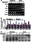
FIGURE 1.
FIGURE 1.
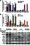
Human iPS cells have an attenuated response to IFN-β. A, IMR-90 and iPS(IMR-90) cells were treated with 1000 IU/ml IFN-β for 15 min, 1 h, and 6 h, and the expression of several ISGs was examined using RT-PCR. B, differences in the induction of ISG expression at 6 h after IFN-β treatment were further quantitated by RT-qPCR. The mRNA expression levels were normalized to actin and gapdh, with gene expression in IMR-90 cells without IFN-β treatment set as “1.” C, protein expression of MX1, MDA5, and RIG-I after IFN-β treatment was analyzed by immunoblotting.
FIGURE 2.
FIGURE 2.
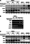
Human ES cells have an attenuated response to IFN-β. A, H9 and H9+BMP4 cells were treated with 1000 IU/ml IFN-β for 15 min, 1 h, and 6 h, and the expression of several ISGs was examined using RT-PCR. B, differences in the induction of ISG expression at 6 h after IFN-β treatment were further quantitated by RT-qPCR. The mRNA expression levels were normalized to actin and gapdh, with the gene expression in H9+BMP4 cells without IFN-β treatment set as “1.” C, protein expression of MX1, MDA5, and RIG-I after 6-h IFN-β treatment was analyzed by immunoblotting.
FIGURE 3.
FIGURE 3.
Expression of IFN signaling molecules in human pluripotent cells. A, lower expression of key type I IFN signaling pathway components in iPS(IMR-90) cells, analyzed by RT-qPCR. B, lower expression of key type I IFN signaling pathway components in H9 cells analyzed by RT-qPCR. The mRNA expression levels were normalized to actin and gapdh. **, p < 0.01; ***, p < 0.001. Error bars, S.E. C, Protein expression of IFN-β signaling molecules in pluripotent and differentiated human cells.
FIGURE 4.
FIGURE 4.
Examination of STAT1 phosphorylation state in pluripotent human cells. A, STAT1 phosphorylation induced by IFN-β is not observed in iPS(IMR-90) cells. Immunoblot analysis of phospho-STAT1(Tyr701) levels in IMR-90 and iPS(IMR-90) cells was performed at different time points after IFN-β treatment. B, immunoblot analysis of phospho-STAT1(Tyr701), phospho-STAT2(Tyr689), and phospho-STAT3(Tyr705) levels in pluripotent and differentiated human cells was done at 6 h after IFN-β treatment.
FIGURE 5.
FIGURE 5.
The attenuated response to IFN-β may be a general feature of pluripotent human cells. A, STAT1 phosphorylation after IFN-β treatment is diminished in H1 and CT2 cells. B, the induction of ISG expression by IFN-β is attenuated in H1 and CT2 cells. C, the increase in MDA5 and RIG-I protein expression in response to IFN-β was not observed in H1e45 or CT2 cells.
FIGURE 6.
FIGURE 6.
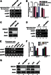
Human pluripotent cells exhibit an elevated expression of SOCS1. A, iPS(IMR-90) cells have significantly higher expression of socs1 than IMR-90 cells, confirmed by RT-qPCR. B and C, elevated SOCS1 protein expression was also observed in iPS(IMR-90) cells. D, RT-PCR reveals that H9 cells have a significantly higher expression of socs1 than H9+BMP4 cells. E, other human embryonic stem cells exhibited higher socs1 expression, as determined by RT-PCR and RT-qPCR. F and G, higher SOCS1 protein expression was also observed in H9, H1, and CT2 cells. The levels of gene expression were normalized to gapdh. ***, p < 0.001. Error bars, S.E.
FIGURE 7.
FIGURE 7.
Reduction of socs1 expression in human iPS cells. A, assessment of socs1 knockdown efficiency in iPS(IMR-90) cells by RT-PCR and RT-qPCR, B and C, immunoblotting analysis, further confirming SOCS1 expression reduction in the knockdown cells. Error bars, S.E.
FIGURE 8.
FIGURE 8.
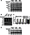
Reduction of socs1 expression leads to a stronger response to IFN-β in human iPS cells. A, immunoblot analysis of phospho-STAT1(Tyr701), phospho-STAT2(Tyr689), and phospho-STAT3(Tyr705) levels in the negative control siRNA- and socs1 siRNAs-treated iPS(IMR-90) cells were performed upon IFN-β treatment. B, cells with lower socs1 levels showed higher induction of ISG expression when treated with IFN-β, which was further quantitated by RT-qPCR, C and D, MDA5 and RIG-I protein expression after IFN-β treatment was also significantly higher in the iPS(IMR-90) cells with reduced socs1 expression. The levels of gene expression were normalized to gapdh. ***, p < 0.001. Error bars, S.E.
FIGURE 9.
FIGURE 9.
Proposed model for the attenuated response to type I IFNs in human pluripotent cells. Although some signaling molecules in the type I IFN response pathway are expressed at lower levels in human pluripotent cells, these cells also exhibit an elevated expression of SOCS1, a potent negative regular of JAK/STAT signaling, compared with differentiated human cells. Together, these may contribute to the diminished STAT1 phosphorylation and a resulting attenuated response to type I IFNs in pluripotent human cells.

References

    1. Isaacs A., Lindenmann J. (1957) Virus interference. I. The interferon. Proc. R. Soc. Lond. B Biol. Sci. 147, 258–267 - PubMed
    1. González-Navajas J. M., Lee J., David M., Raz E. (2012) Immunomodulatory functions of type I interferons. Nat. Rev. Immunol. 12, 125–135 - PMC - PubMed
    1. Platanias L. C. (2005) Mechanisms of type I- and type II-interferon-mediated signalling. Nat. Rev. Immunol. 5, 375–386 - PubMed
    1. Shultz L. D., Rajan T. V., Greiner D. L. (1997) Severe defects in immunity and hematopoiesis caused by SHP-1 protein-tyrosine phosphatase deficiency. Trends Biotechnol. 15, 302–307 - PubMed
    1. Zhang J., Somani A. K., Siminovitch K. A. (2000) Roles of the SHP-1 tyrosine phosphatase in the negative regulation of cell signalling. Semin. Immunol. 12, 361–378 - PubMed

Publication types

MeSH terms