. 2013 Jun 14;288(24):17759-68.
doi: 10.1074/jbc.M113.477133. Epub 2013 Apr 17.

Protein kinase C δ increases Kruppel-like factor 4 protein, which drives involucrin gene transcription in differentiating keratinocytes

Affiliations

Protein kinase C δ increases Kruppel-like factor 4 protein, which drives involucrin gene transcription in differentiating keratinocytes

Yap Ching Chew et al. J Biol Chem. .

Abstract

KLF4 is a member of the Kruppel-like factor family of transcriptional regulators. KLF4 has been shown to be required for normal terminal differentiation of keratinocytes, but the molecular mechanism whereby KLF4 regulates genes associated with the differentiation process has not been studied. In the present study, we explore the impact of KLF4 on expression of involucrin, a gene that is specifically expressed in differentiated keratinocytes. KLF4 overexpression and knockdown studies show that involucrin mRNA and protein level correlates directly with KLF4 level. Moreover, studies of mutant KLF4 proteins indicate that transcriptionally inactive forms do not increase involucrin expression. PKCδ is a regulator of keratinocyte differentiation that increases expression of differentiation-associated target genes, including involucrin. Overexpression of KLF4 augments the PKCδ-dependent increase in involucrin expression, whereas KLF4 knockdown attenuates this response. The KLF4 induction of human involucrin (hINV) promoter activity is mediated via KLF4 binding to a GC-rich element located in the hINV promoter distal regulatory region, a region of the promoter required for in vivo involucrin expression. Mutation of the GC-rich element, an adjacent AP1 factor binding site, or both sites severely attenuates the response. Moreover, loss of KLF4 in an epidermal equivalent model of differentiation results in loss of hINV expression. These studies suggest that KLF4 is part of a multiprotein complex that interacts that the hINV promoter distal regulatory region to drive differentiation-dependent hINV gene expression in epidermis.

Keywords: Cell Differentiation; Gene Expression; Keratinocytes; Kruppel-like Factor (KLF); Protein Kinase C (PKC).


Figures
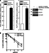
FIGURE 1.
FIGURE 1.
PKCδ controls KLF4 mRNA and protein level. A, keratinocytes (KERn) were infected with 15 MOI of Ad5-EV or Ad5-PKCδ, and after 24 h, extracts were prepared for detection of PKCδ and KLF4 mRNA and protein. The mRNA abundance values are mean ± S.D., n = 3. The asterisk indicates a significant increase over control as determined by Student's t test, p < 0.005. For immunoblot, cells were treated with adenovirus as above, and after 24 h, extracts were prepared for detection of the indicated epitopes. B, PKCδ does not alter KLF4 mRNA half-life. Keratinocytes were infected with 15 MOI of Ad5-EV or Ad5-PKCδ and maintained for 24 h. Actinomycin D was then added to the cultures, and mRNA was harvested at 0–4 h after the addition of 5 μg/ml actinomycin. KLF4 mRNA was detected by quantitative RT-PCR. The RNA decay data are presented as a log-linear plot to determine first-order decay constant.
FIGURE 2.
FIGURE 2.
KLF4 expression increases hINV expression. A, keratinocytes (KERn) were infected with 15 MOI of tAd5-EV or tAd5-hKLF4. After 48 h, extracts were prepared for detection of KLF4 and hINV protein. B, KLF4 increases hINV mRNA level. Keratinocytes were infected with the indicated virus, and after 24 h, RNA was harvested for detection of KLF4 mRNA by quantitative RT-PCR. The values are the mean ± S.D., n = 3. The asterisk indicates a significant increase over control as determined by Student's t test, p < 0.005. Similar results were observed in each of three experiments. C, keratinocytes were transfected with 2 μg of pINV-2473 luciferase reporter plasmid in the presence of 1 μg of empty vector or the indicated KLF4 expression vector. At 24 h, the cells were harvested, and extracts were assayed for luciferase activity. The asterisk indicates a significant increase (n = 3) as determined by Student's t test, p < 0.005. Wild-type and mutant KLF4 expression was monitored by immunoblot and normalized to the level of β-actin. hKLF4(335–470) could not be monitored because the antibody epitope is deleted from this mutant. D, keratinocytes were electroporated with 3 μg of the indicated siRNA, and after 48 h, cells were transfected with 4 μg of luciferase reporter plasmid. After an additional 24 h, extracts were prepared for luciferase activity assay and immunoblot. The values are mean ± S.D., n = 3. The asterisk indicates a significant reduction in luciferase activity, p < 0.005. Similar results were observed in each of three experiments. Identical results were observed using several KLF4 siRNA, indicating that the observed responses are not due to off-target effects.
FIGURE 3.
FIGURE 3.
KLF4 is required for PKCδ-induced hINV expression. A, the PKCδ-dependent increase in hINV mRNA level requires KLF4. Keratinocytes (KERn) were electroporated with 3 μg of the indicated siRNA, and at 36 h, cells were infected with 15 MOI of Ad5-EV or Ad5-PKCδ. After an additional 36 h, the cells were harvested for assay of hINV mRNA and protein. Similar results were observed in each of three experiments. Moreover, identical results were observed using several KLF4-specific siRNA, indicating that the observed responses are specific. B and C, the TPA-dependent increase in hINV mRNA level requires KLF4. Keratinocytes were electroporated with 3 μg of the indicated siRNA. After 36 h, the cells were treated in the presence or absence of TPA (50 ng/ml) for an additional 36 h. Extracts were then prepared for detection hINV mRNA and luciferase activity. Similar results were observed in each of three experiments. Identical results were observed using several KLF4 siRNA, indicating that the observed responses are not due to off-target effects. D, KLF4 augments the PKCδ-dependent increase in hINV promoter activity. Keratinocytes were transfected with 2 μg of hINV luciferase reporter plasmid, 1 μg of KLF4 expression vector, and 1 μg of PKCδ expression vector. After 24 h, the cells were harvested, and extracts were assayed for luciferase activity. E, KLF4 augments the TPA-dependent increase in hINV promoter activity. Keratinocytes were transfected with 2 μg of involucrin luciferase reporter plasmid in the presence or absence of 1 μg of KLF4 expression vector. After 24 h, the cells were treated with or without TPA (50 ng/ml) for 24 h prior to assay for luciferase activity. In all panels, the values are the mean ± S.D. (n = 3), and the asterisks indicate a significant increase or decrease as determined using the Student's t test, p < 0.005. Similar results were obtained in each of three independent experiments.
FIGURE 4.
FIGURE 4.
KLF4 activation of hINV promoter requires the DRR. A, the hINV promoter upstream regulatory region showing functionally important (–39, 50, 52, 53) AP1 (AP1-1 and AP1-5) and GC-rich (Sp1 binding) (22, 37, 38) response elements. The two biologically important AP1 sites present within the upstream regulatory region are indicated (AP1-5 and AP1-1), as is the Sp1 site. The distances are in nucleotides relative to the transcription start site. B, KLF4 regulation of hINV promoter activity requires the DRR. Keratinocytes were transfected with 2 μg of the indicated hINV reporter plasmid and 1 μg of empty expression vector or KLF4 expression vector for 24 h prior to harvest and assay for luciferase activity. In all cases, the values are the mean ± S.D. (n = 3), and the asterisk indicates a significant increase or decrease, p < 0.005. Similar results were observed in three independent experiments.
FIGURE 5.
FIGURE 5.
KLF4 activation of hINV promoter activity requires the DRR AP1 and GC-rich response elements. A, schematic showing key regulatory elements in the hINV promoter. pINV-2473 is the full-length promoter. pINV(−2473/−2088) is a construct in which the DRR region (nucleotides −2473/−2088) is linked to the hINV minimal promoter (−41/−1) The dashed line indicates the fusion. The functionally important AP1 (AP1-1 and AP1-5) and GC-rich element are indicated. The distances are in nucleotides relative to the transcription start site. B and C, keratinocytes (KERn) were transfected with 2 μg of the indicated reporter plasmid and 1 μg of empty vector or hKLF4-expression vector, and after 24 h, the cells were harvested, and extracts were prepared for luciferase activity assay. The values are mean ± S.D., n = 3. In all cases, the asterisk indicates a significant reduction in luciferase activity as determined using the Student's t test, p < 0.005.
FIGURE 6.
FIGURE 6.
KLF4 interacts with AP1-5/GC-rich response element in the hINV promoter DRR. A and B, keratinocytes (KERn) were infected with 15 MOI of tAd5-EV or tAd5-hKLF4. After 48 h, cells were prepared for ChIP assay. ChIP was performed as described under “Experimental Procedures” using hINV promoter-derived PCR primers encoding the indicated range of nucleotides. In all panels, the values are the mean ± S.D. (n = 3), and the asterisk indicates a significant increase or decrease as determined using the Student's t test, p < 0.005.
FIGURE 7.
FIGURE 7.
KLF4 is required for hINV expression during keratinocyte differentiation. A and C, keratinocytes (KERn) were electroporated with control or KLF4 siRNA and seeded into Millicell wells to form epidermal equivalents. After 4 days, the epidermal equivalents were harvested, and extracts were prepared for assay of KLF4 and hINV level. B, epidermal equivalents were sectioned and stained with hematoxylin/eosin to assess differentiation status. Bar = 100 μm, m indicates the membrane, and c indicates the cornified layer. The arrows indicate nuclei that are retained in cells in the suprabasal layers of the KLF4 siRNA-treated epidermal equivalent. Identical results were obtained with three independent sets of control and KLF4 siRNA. In all cases, the values are the mean ± S.D. (n = 3), and the asterisks indicate a significant increase or decrease, p < 0.005.

References

    1. Kaczynski J., Cook T., Urrutia R. (2003) Sp1- and Krüppel-like transcription factors. Genome Biol. 4, 206. - PMC - PubMed
    1. Swamynathan S. K., Davis J., Piatigorsky J. (2008) Identification of candidate KLF4 target genes reveals the molecular basis of the diverse regulatory roles of KLF4 in the mouse cornea. Invest. Ophthalmol. Vis. Sci. 49, 3360–3370 - PMC - PubMed
    1. Patel S., Xi Z. F., Seo E. Y., McGaughey D., Segre J. A. (2006) Klf4 and corticosteroids activate an overlapping set of transcriptional targets to accelerate in utero epidermal barrier acquisition. Proc. Natl. Acad. Sci. U.S.A. 103, 18668–18673 - PMC - PubMed
    1. Chen X., Whitney E. M., Gao S. Y., Yang V. W. (2003) Transcriptional profiling of Krüppel-like factor 4 reveals a function in cell cycle regulation and epithelial differentiation. J. Mol. Biol. 326, 665–677 - PMC - PubMed
    1. Whitney E. M., Ghaleb A. M., Chen X., Yang V. W. (2006) Transcriptional profiling of the cell cycle checkpoint gene Krüppel-like factor 4 reveals a global inhibitory function in macromolecular biosynthesis. Gene Expr. 13, 85–96 - PMC - PubMed

Publication types

LinkOut - more resources