. 2013 Apr 19;340(6130):337-41.
doi: 10.1126/science.1231391.

Multicompartment mesoporous silica nanoparticles with branched shapes: an epitaxial growth mechanism

Affiliations

Multicompartment mesoporous silica nanoparticles with branched shapes: an epitaxial growth mechanism

Teeraporn Suteewong et al. Science. .

Abstract

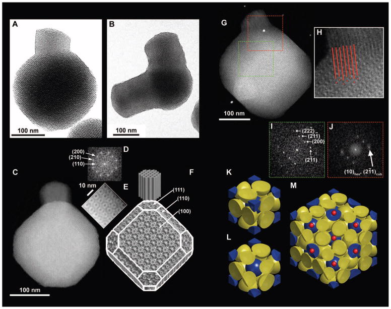
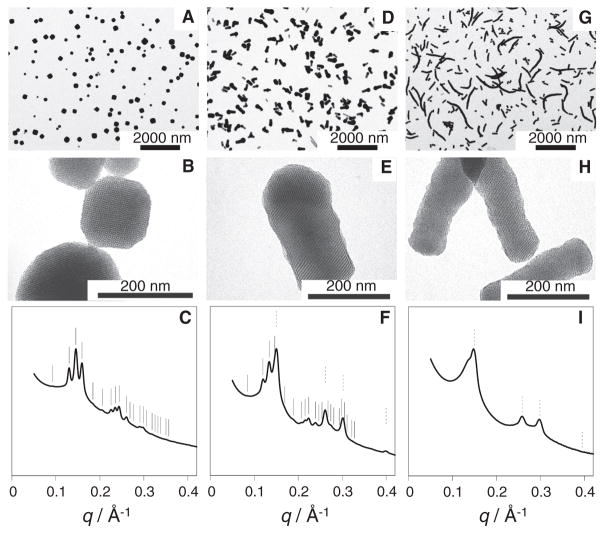
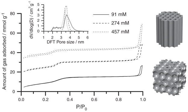
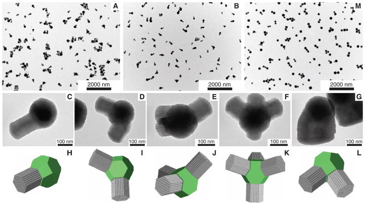
Mesoporous nanomaterials have attracted widespread interest because of their structural versatility for applications including catalysis, separation, and nanomedicine. We report a one-pot synthesis method for a class of mesoporous silica nanoparticles (MSNs) containing both cubic and hexagonally structured compartments within one particle. These multicompartment MSNs (mc-MSNs) consist of a core with cage-like cubic mesoporous morphology and up to four branches with hexagonally packed cylindrical mesopores epitaxially growing out of the cubic core vertices. The extent of cylindrical mesostructure growth can be controlled via a single additive in the synthesis. Results suggest a path toward high levels of architectural complexity in locally amorphous, mesostructured nanoparticles, which could enable tuning of different pore environments of the same particle for specific chemistries in catalysis or drug delivery.


Figures

Fig. 1
Fig. 1. Geometrical description of mc-MSNs
(A) BF-TEM image of a branch with hexagonal pore structure emanating from one corner of a core with cubic pore structure. (B) BF-TEM image showing two branches emanating from two corners of the core. (C) HAADF-STEM image of (100) projection of the core. (D) FFT of the entire cubic core part of the image in (C). (E) Magnified image of the top right edge of the particle in (C). (F) Schematics of a model mc-MSN. Miller indices for some of the representative facets are provided as a visual guide. (G) HAADF-STEM image of a mc-MSN exhibiting the (110) projection of the core. (H) Magnified image of the connecting region between the core and branch in (G), with red lines and dots indicating projected vacancy positions to demonstrate the structural registry/epitaxy. (I) FFT of the cubic core region in (G) (green box). (J) FFT of the connected region in (G) (red box). (K to M) Model visualization of the epitasial relationship of mesopores at the interface of (111) Pmn cubic and (0001) P6mm hexagonal planes. (K) Unit cell of the Pmn cage-like structure exhibiting body-centered cubic lattice micelles (blue spheres) and pairs of micelles on the faces (yellow spheres). (L) (111) plane cut of a single unit cell. (M) (111) plane cut of a 2 × 2 × 2 lattice, additionally showing the positions of expected hexagonal channels as small red dots.
Fig. 2
Fig. 2. TEM images and SAXS patterns of aminated MSNs synthesized with varying [EtOAc]
(A, D, and G) Low-magnification TEM images of MSNs prepared from (A) 91 mM, (B) 274 mM, and (C) 457 mM EtOAc. (B, E, and H) Higher-magnification TEM images of the MSNs in (A), (D), and (G). (C, F, and I) The corresponding SAXS patterns (q denotes the scattering vector magnitude, defined as q = 4π sin θ/λ, where 2θ is the total scattering angle and λ is the x-ray wavelength). In the SAXS patterns, expected peak positions from cubic and hexagonal lattices are indicated by solid and dotted lines, respectively. Data for 91 mM EtOAc are adapted from (19).
Fig. 3
Fig. 3. Nitrogen sorption isotherms of MSNs synthesized with varying [EtOAc]
Isotherms for mc-MSNs prepared from 274 mM and 457 mM EtOAc are offset along the y axis by 15 and 30 mmol/g, respectively (P/P0 denotes the nitrogen partial pressure). The inset shows pore size distributions obtained from NLDFT calculations based on the respective absorption branches. The models on the right provide a direct comparison of the pore structures of hexagonal and cubic lattices.
Fig. 4
Fig. 4. TEM images and models of mc-MSNs with complex mesostructures
(A and B) Low-magnification TEM images of mc-MSNs prepared from (A) 137 mM and (B) 183 mM EtOAc. (C to G) High-magnification TEM images of mc-MSNs in the 183 mM EtOAc batch with (C) one arm, (D) two arms, (E) three arms, (F) four arms, and (G) two arms merged into one. (H to L) Models corresponding to the images in (C) to (G). Cores are shown as green truncated cubes; branches are represented as gray columns. (M) Low-magnification TEM image of mc-MSNs where additional silane precursors (one-half of the initial amount) were injected after 30 min of aging time (21).

References

    1. Yanagisawa T, Shimizu T, Kuroda K, Kato C. Bull Chem Soc Jpn. 1990;63:988.
    1. Kresge CT, Leonowicz ME, Roth WJ, Vartuli JC, Beck JS. Nature. 1992;359:710.
    1. Hoffmann F, Cornelius M, Morell J, Fröba M. Angew Chem Int Ed. 2006;45:3216. - PubMed
    1. Xiao CH, Fujita N, Miyasaka K, Sakamoto Y, Terasaki O. Nature. 2012;487:349. - PubMed
    1. Vallet-Regí M, Balas F, Arcos D. Angew Chem Int Ed. 2007;46:7548. - PubMed

Publication types

Substances

LinkOut - more resources