Clinician-performed Beside Ultrasound for the Diagnosis of Traumatic Pneumothorax
- PMID: 23599841
- PMCID: PMC3628453
- DOI: 10.5811/westjem.2012.12.12663
Clinician-performed Beside Ultrasound for the Diagnosis of Traumatic Pneumothorax
Abstract
Introduction: Prior studies have reported conflicting results regarding the utility of ultrasound in the diagnosis of traumatic pneumothorax (PTX) because they have used sonologists with extensive experience. This study evaluates the characteristics of ultrasound for PTX for a large cohort of trauma and emergency physicians.
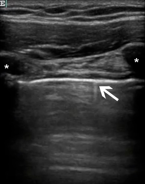
Methods: This was a prospective, observational study on a convenience sample of patients presenting to a trauma center who had a thoracic ultrasound (TUS) evaluation for PTX performed after the Focused Assessment with Sonography for Trauma exam. Sonologists recorded their findings prior to any other diagnostic studies. The results of TUS were compared to one or more of the following: chest computed tomography, escape of air on chest tube insertion, or supine chest radiography followed by clinical observation.
Results: There were 549 patients enrolled. The median injury severity score of the patients was 5 (inter-quartile range [IQR] 1-14); 36 different sonologists performed TUS. Forty-seven of the 549 patients had traumatic PTX, for an incidence of 9%. TUS correctly identified 27/47 patients with PTX for a sensitivity of 57% (confidence interval [CI] 42-72%). There were 3 false positive cases of TUS for a specificity of 99% (CI 98%-100%). A "wet" chest radiograph reading done in the trauma bay showed a sensitivity of 40% (CI 23-59) and a specificity of 100% (99-100).
Conclusion: In a large heterogenous group of clinicians who typically care for trauma patients, the sonographic evaluation for pneumothorax was as accurate as supine chest radiography. Thoracic ultrasound may be helpful in the initial evaluation of patients with truncal trauma.
Figures

References
-
- Kirkpatrick AW, Ng AKT, Dulchavsky SA, et al. Sonographic Diagnosis of a Pneumothorax Inapparent on Plain Radiography: Confirmation by Computed Tomography. The Journal of Trauma: Injury, Infection, and Critical Care. 2001 Apr;50(4):750–752. - PubMed
-
- Lichtenstein DA, Menu Y. A Bedside Ultrasound Sign Ruling Out Pneumothorax in the Critically III: Lung Sliding. Chest. 1995 Nov 1;108(5):1345–1348. - PubMed
-
- Mowery NT, Gunter OL, Collier BR, et al. Practice management guidelines for management of hemothorax and occult pneumothorax. J Trauma. 2011 Feb;70(2):510–518. - PubMed
-
- Lichtenstein D, Mezière G, Biderman P, et al. The comet-tail artifact: an ultrasound sign ruling out pneumothorax. Intensive Care Medicine. 1999 Apr;25(4):383–388. - PubMed
-
- Dulchavsky SA, Schwarz KL, Kirkpatrick aW, et al. Prospective evaluation of thoracic ultrasound in the detection of pneumothorax. The Journal of Trauma. 2001 Feb;50(2):201–205. - PubMed