. 2013 Sep 1;22(17):2347-55.
doi: 10.1089/scd.2012.0415. Epub 2013 May 24.

Not all MSCs can act as pericytes: functional in vitro assays to distinguish pericytes from other mesenchymal stem cells in angiogenesis

Affiliations

Not all MSCs can act as pericytes: functional in vitro assays to distinguish pericytes from other mesenchymal stem cells in angiogenesis

Anna Blocki et al. Stem Cells Dev. .

Abstract

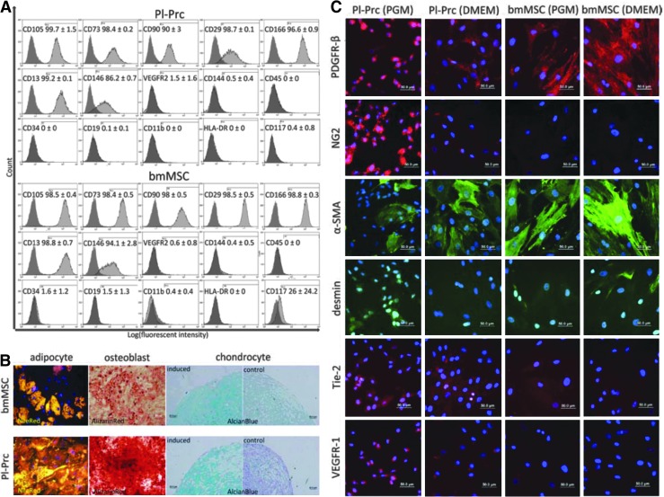
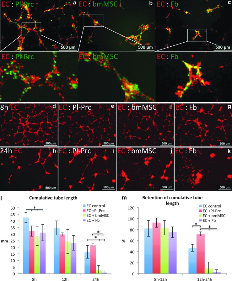
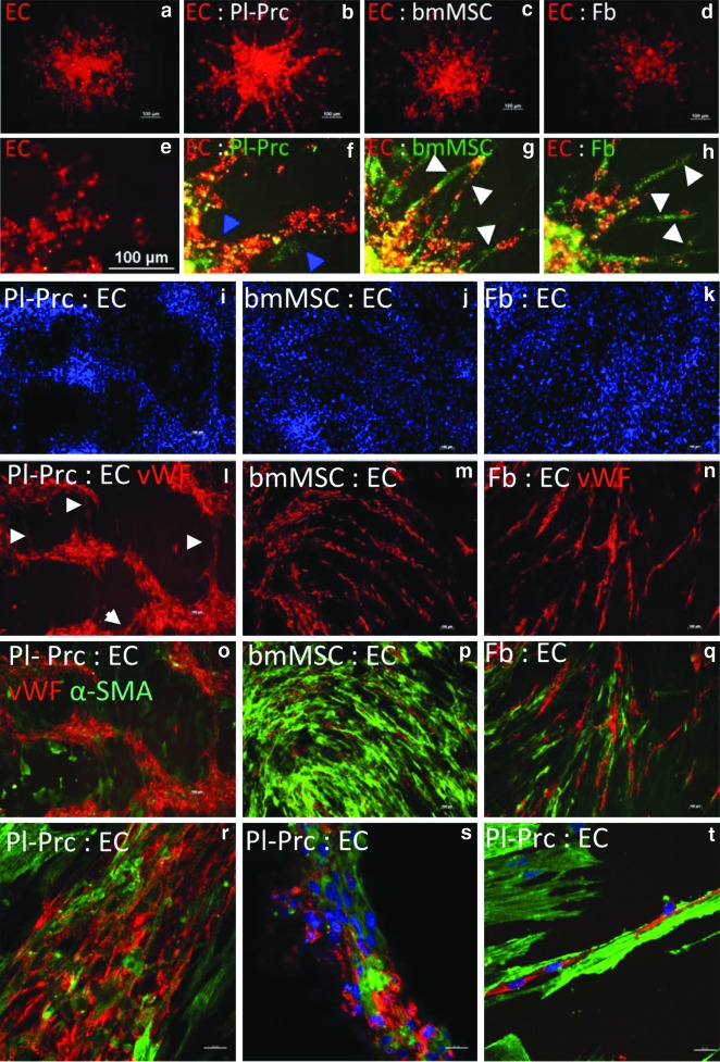
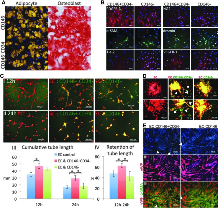
Pericytes play a crucial role in angiogenesis and vascular maintenance. They can be readily identified in vivo and isolated as CD146(+)CD34(-) cells from various tissues. Whether these and other markers reliably identify pericytes in vitro is unclear. CD146(+)CD34(-) selected cells exhibit multilineage potential. Thus, their perivascular location might represent a stem cell niche. This has spurred assumptions that not only all pericytes are mesenchymal stromal cells (MSCs), but also that all MSCs can act as pericytes. Considering this hypothesis, we developed functional assays by confronting test cells with endothelial cultures based on matrigel assay, spheroid sprouting, and cord formation. We calibrated these assays first with commercial cell lines [CD146(+)CD34(-) placenta-derived pericytes (Pl-Prc), bone marrow (bm)MSCs and fibroblasts]. We then functionally compared the angiogenic abilities of CD146(+)CD34(-)selected bmMSCs with CD146(-) selected bmMSCs from fresh human bm aspirates. We show here that only CD146(+)CD34(-) selected Pl-Prc and CD146(+)CD34(-) selected bmMSCs maintain endothelial tubular networks on matrigel and improve endothelial sprout morphology. CD146(-) selected bmMSCs neither showed these abilities, nor did they attain pericyte function despite progressive CD146 expression once passaged. Thus, cell culture conditions appear to influence expression of this and other reported pericyte markers significantly without correlation to function. The newly developed assays, therefore, promise to close a gap in the in vitro identification of pericytes via function. Indeed, our functional data suggest that pericytes represent a subpopulation of MSCs in bm with a specialized role in vascular biology. However, these functions are not inherent to all MSCs.


Figures

FIG. 1.
FIG. 1.
Pl-Prc are MSCs but express additional markers when propagated in PGM. (A) Pl-Prc and bmMSCs (n=3) share a common MSC marker profile as indicated by MSC marker (CD105 through CD13), also CD146 (used for pericyte isolation), and absence of endothelial/hematopoietic markers (CD144 through CD117). (B) Pl-Prc and bmMSCs (n=3) were induced to differentiate into adipocytes, osteoblasts, and chondrocytes. Lipid droplets (adipocyte) were stained with nile red (gold), calcification (osteoblast) with alizarin red (red) and proteoglycans (chondrocyte) with alcian blue (blue), respectively. (C) Immunocytochemical detection of pericytic markers. Presence of PDGFR-β and α-SMA is independent from culture medium; however, NG2, desmin, Tie-2 and VEGFR-1 are more strongly expressed in Pl-Prc cultured in PGM. (A–C) Results are representatives of three independent studies. α-SMA, α-smooth muscle actin; bmMSC, bone marrow mesenchymal stromal cells; MSCs, mesenchymal stromal cells; PDGFR-β, platelet derived growth factor β; PGM, pericyte growth media; Pl-Prc, placenta-derived pericytes.
FIG. 2.
FIG. 2.
Only Pl-Prc stabilize endothelial networks. Coculture of HUVEC with Pl-Prc, bmMSCs or IMR90s (Fb) on matrigel (n≥3). (a–c) In mixed cultures all mesenchymal cells (green label) colocalized with the endothelial network (red label). Coculture at a ratio of 20:1 at 8 h (d–g) and 24 h (h–k) demonstrate formation of EC network (only red labeled EC shown) and decay at 24 h. In the presence of Pl-Prc, the decay is slowed. (l) Quantitation of cumulative tube length (*P<0.02) and (m) retention of cumulative tube length at various time intervals (*P≤0.01) demonstrate that only Pl-Prc can significantly retain integrity of endothelial tubules over 24 h. Results are displayed as average±standard deviation. Results are representative of three independent studies. EC, endothelial cells; HUVECs, human umbilical vein endothelial cells.
FIG. 3.
FIG. 3.
Only Pl-Prc improve sprout integrity and move with endothelial cells to form cords in coculture. EC (red label) were cocultured with Pl-Prc, bmMSCs, IMR90s (Fb) (green label) to form spheroids (n=24), which were introduced into collagen I gels to sprout. (a–d) only red labeled EC are shown. (a) EC cultures show spontaneous formation of loosely arranged sprouts. (b) In the presence of Pl-Prc, formed sprouts are broader and have a smooth compact morphology. (c, d) In the presence of bmMSCs and Fb, sprouts appear thin and discontinuous. (e–h) Close-ups reveal that Pl-Prc colocalize with EC (blue arrowheads), while bmMSCs and Fb segregate from EC or bridge detached ECs (white arrowheads). (i–t) In a cord forming coculture assay EC were seeded on top of confluent Pl-Prc, bmMSCs, or Fb (n≥3). While bmMSCs and Fb support cord formation of single-cell thickness (m, n), only Pl-Prc show formation of cords of various thicknesses with interconnections (l, white arrowheads). (r–t) Confocal analyses reveal that Pl-Prc incorporate into formed cords and are aligned parallel to cord direction. (t) α-SMA fibers are more pronounced in Pl-Prc contributing to cords and are aligned in the same direction. Results are representatives of three independent studies.
FIG. 4.
FIG. 4.
CD146+CD34 selected but not CD146 selected bmMSCs stabilize endothelial networks and improve endothelial sprout integrity. CD146+CD34 cells and CD146 cells from the same bm specimens were subjected to: (A) Induction into adipocytes and osteoblasts. Lipid droplets were stained with nile red (gold) and calcification was stained with alizarin red (red) in the respective cultures. Cells were confirmed to be MSCs. (B) Immunocytochemistry of pericyte-related markers. Cells share a very similar marker profile of PDGFR-β, α-SMA, NG2, desmin, Tie-2, and VEGFR-1. (C) Coculture with HUVEC (1:20) on matrigel (n≥3). (I, II) Superimposition of signals for EC (red) and cocultured bm-derived cells (green) at 12 and 24 h. (III) Quantitation of cumulative tube length (*P<0.01). (IV) Quantitation of retention of cumulative tube length (*P<0.01). Quantitative results are displayed as average value±standard deviation. CD146+CD34 cells stabilize endothelial network significantly. (D) Mixed spheroid sprouting assay. EC (red label) were introduced as spheroids (n=24) with bm derived cells (green label) into collagen I gels to sprout. (I, II) Results using cells from two independent samples are displayed. (E) Mono-layer coculture assay. (I) EC were seeded on top of CD146+CD34 cells and CD146 cells at 1:4 in media containing VEGF and cultured for 3 days. Methanol fixed cocultures were immunostained for vWF (red, EC maker) (II) and α-SMA (green, mesenchymal cell marker) (III), and nuclei were stained with DAPI (blue) (n=2). CD146+CD34 cells move slightly more towards formed endothelial cords resulting in areas in between cords with less DAPI and no α-SMA (white arrowheads). Results are representatives of studies performed with three different bm samples.

References

    1. Sims DE. The pericyte—a review. Tissue Cell. 1986;18:153–174. - PubMed
    1. Armulik A. Genové G. Betsholtz C. Pericytes: developmental, physiological, and pathological perspectives, problems, and promises. Dev Cell. 2011;21:193–215. - PubMed
    1. Song S. Ewald AJ. Stallcup W. Werb Z. Bergers G. PDGFRbeta+ perivascular progenitor cells in tumours regulate pericyte differentiation and vascular survival. Nat Cell Biol. 2005;7:870–879. - PMC - PubMed
    1. Darland DC. D'Amore PA. TGF beta is required for the formation of capillary-like structures in three-dimensional cocultures of 10T1/2 and endothelial cells. Angiogenesis. 2001;4:11–20. - PubMed
    1. Dar A. Domev H. Ben-Yosef O. Tzukerman M. Zeevi-Levin N. Novak A, et al. Multipotent vasculogenic pericytes from human pluripotent stem cells promote recovery of murine ischemic limb. Circulation. 2012;125:87–99. - PubMed

Publication types