. 2013 Jun;172(3):417-26.
doi: 10.1111/cei.12062.

Ethyl pyruvate significantly inhibits tumour necrosis factor-α, interleukin-1β and high mobility group box 1 releasing and attenuates sodium taurocholate-induced severe acute pancreatitis associated with acute lung injury

Affiliations

Ethyl pyruvate significantly inhibits tumour necrosis factor-α, interleukin-1β and high mobility group box 1 releasing and attenuates sodium taurocholate-induced severe acute pancreatitis associated with acute lung injury

Z-G Luan et al. Clin Exp Immunol. 2013 Jun.

Abstract

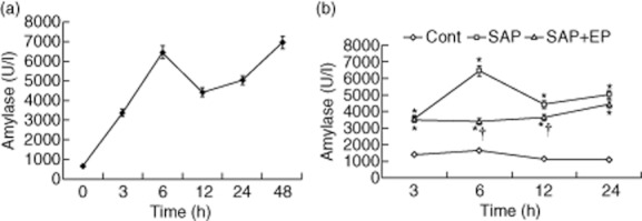
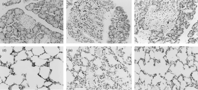
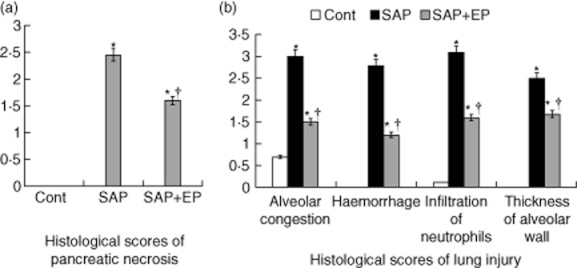
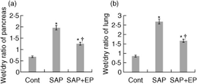
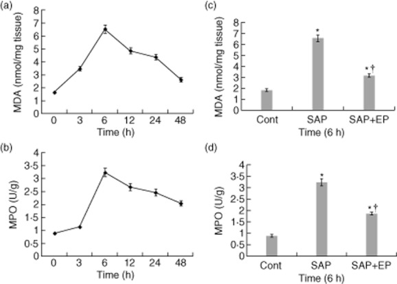
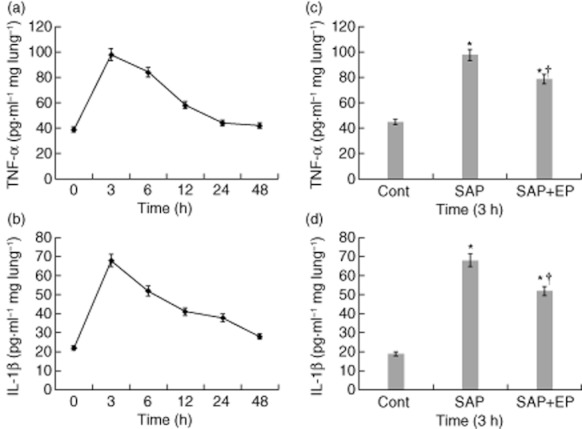
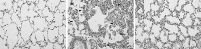
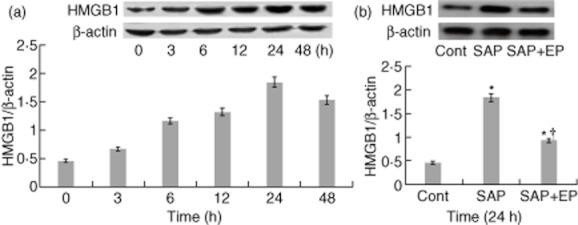
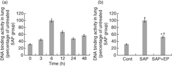
In this study, we examined the effect of ethyl pyruvate (EP) on pulmonary inflammation in rats with severe pancreatitis-associated acute lung injury (ALI). Severe acute pancreatitis (SAP) was induced in rats by the retrograde injection of 5% sodium taurocholate into the pancreatic duct. Rats were randomly divided into the following experimental groups: control group, SAP group and EP-treated group. The tissue specimens were harvested for morphological studies, Streptavidin-peroxidase immunohistochemistry examination. Pancreatic or lung tissue oedema was evaluated by tissue water content. Serum amylase and lung tissue malondialdehyde (MDA) and myeloperoxidase (MPO) were measured. Meanwhile, the nuclear factor-κB (NF-κB) activation, tumour necrosis factor-α (TNF-α), interleukin-1β (IL-1β) levels and HMGB1 protein expression levels in the lung were studied. In the present study, we demonstrated that treatment with EP after SAP was associated with a reduction in the severity of SAP and lung injury. Treatment with EP significantly decreased the expression of TNF-α, IL-1β, HMGB1 and ameliorated MDA concentration, MPO activity in the lung in SAP rats. Compared to SAP group, administration of EP prevented pancreatitis-induced increases in nuclear translocation of NF-κB in the lung. Similarly, treatment with EP significantly decreased the accumulation of neutrophils and markedly reduced the enhanced lung permeability. In conclusion, these results demonstrate that EP might play a therapeutic role in pulmonary inflammation in this SAP model.


Figures

Fig. 1
Fig. 1
Serum amylase levels are measured in rats of three groups. Male Wistar rats were induced to severe acute pancreatitis (SAP), and serum was collected at different time-points and assayed for amylase levels. Data are shown as the means ± standard error (n = 12 rats per time-point in per subgroup). Lozenges: control group; squares: SAP group; triangles: EP-treated group. * P < 0·05 versus control group and † P < 0·05 versus SAP group, as tested by one-way analysis of variance (anova). The level at zero time means the point of first injection of sodium taurocholate. Cont: control.
Fig. 2
Fig. 2
Morphological changes of pancreas and lung stained with haematoxylin and eosin (H&E). (a) No histological alteration of pancreas was observed in the pancreas collected from the control group. (b) Histological examination [at 24 h after severe acute pancreatitis (SAP)] of pancreatic sections from pancreatitic rats revealed oedema and acinar cell necrosis as well as inflammatory cells infiltration and an important alteration of the pancreas was also present. (c) Significantly less histological alteration of the pancreas tissue was observed in pancreatitic rats, which received ethyl pyruvate (EP) treatment. Representative H&E-stained section of lung was examined by light microscopy in control rats (d) and in pancreatitic rats (e), and in pancreatitic rats, which received EP treatment (f). Original magnification: ×400. Figure is representative of at least three experiments performed on different experimental days.
Fig. 3
Fig. 3
Histological scores of pancreatic necrosis and lung injury. Pancreas and lung were removed 24 h after induction of severe acute pancreatitis (SAP), and haematoxylin and eosin (H&E) staining was performed. (a) The extent of pancreatic acinar cell necrosis was quantitated morphometrically by an observer who was not aware of the sample identity. (b) Changes in lung histology score. The identified histological changes included congestion, oedema, inflammatory cells and haemorrhaging 24 h after induction of SAP. Histological scores were improved significantly in rats treated with EP compared with pancreatitic rats. *P < 0·05 versus control group and †P < 0·05 versus SAP group, as tested by one-way analysis of variance (anova). Cont: control.
Fig. 4
Fig. 4
Wet/dry weight ratio of pancreatic and lung tissues in rats with severe acute pancreatitis (SAP). (a) The pancreas wet : dry weight (W/D) ratio was obtained to evaluate pancreas oedema. The W/D ratio in pancreas was determined 12 h after the induction of SAP (n = 12 for each group). (b) Effects of ethyl pyruvate (EP) on the lung W/D ratio of taurocholate-treated rats. The lung W/D ratio 12 h following taurocholate administration was shown for each group (n = 12 for each group). The data are expressed as the means ± standard error. *P < 0·05 versus control group and †P < 0·05 versus SAP group, as tested by one-way analysis of variance (anova). Cont: control.
Fig. 5
Fig. 5
Myeloperoxidase (MPO) activity and malondialdehyde (MDA) levels in the lung of pancreatitic rats. Sample was obtained after induction of severe acute pancreatitis (SAP) at the corresponding time-points. (a,c) Effects of ethyl pyruvate (EP) on MDA concentration in the lung of rats with SAP. Pulmonary MDA concentration was significantly lower in EP-treated rats than in SAP rats at 6 h after the induction of SAP. (b,d) Lung neutrophil infiltration as measured by MPO activity. MPO activity was increased significantly in the lung from taurocholate-treated rats in comparison to control rats. The treatment with EP reduced the taurocholate-induced increase of MPO activity significantly in the lung at 6 h after the induction of SAP. Bars represent the means ± standard error of 12 rats. *P < 0·05 versus control group and †P < 0·05 versus SAP group, as tested by one-way analysis of variance (anova). Cont: control.
Fig. 6
Fig. 6
Effects of ethyl pyruvate (EP) treatment on pulmonary tumour necrosis factor (TNF)-α and interleukin (IL)-1β production in pancreatitic rats. (a,b) TNF-α and IL-1β protein of lung in rats was examined by enzyme-linked immunosorbent assay (ELISA) at the designated time-points after SAP. (c, d) At 3 h after the induction of severe acute pancreatitis (SAP), levels of TNF-α and IL-1β were increased compared with control rats. While SAP rats receiving the treatment with EP had a significant reduction in pulmonary TNF-α and IL-1β expression compared with animals receiving vehicle. The number of rats in each group at every time-point was 12. Values are shown means ± standard error. *P < 0·05 versus control group and †P < 0·05 versus untreated SAP group, as tested by one-way analysis of variance (anova). Cont: control.
Fig. 7
Fig. 7
Lung immunohistochemical localization of high mobility group box 1 (HMGB1) is depicted. Immunohistochemical analysis was used to detect HMGB1 protein in lung sections obtained 24 h after severe acute pancreatitis (SAP). Representative specimens from the control group, SAP group and ethyl pyruvate (EP)-treated group are presented. (a) Immunohistochemical stain of HMGB1 from sections of control lung. (b) Staining of lung tissue sections obtained from SAP group with anti-HMGB1 antibody show an intense positive staining in the endothelial cells (white arrows), macrophages and neutrophils (black arrows). (c) The degree of lung staining for HMGB1 was reduced markedly in tissue section obtained from the EP-treated group. The arrows in (b) indicate cells staining positive for HMGB1. All photographs are at ×200 magnification. Images are representative lung sections from 12 rats per group.
Fig. 8
Fig. 8
Changes in high mobility group box 1 (HMGB1) protein expression in lung tissue after induction of severe acute pancreatitis (SAP) in rats. (a) The expression of HMGB1 in the lung was detected by Western blot at the designated time-points after SAP. Results show the HMGB1/β-actin ratio from Western blots performed at each time-point. Blot shown is representative of three experiments with similar results. (b) Effect of treatment with EP on pulmonary expression of HMGB1 at 24 h after SAP. Pulmonary expressions of HMGB1 and β-actin determined by Western blot analysis in the control group, SAP group and ethyl pyruvate (EP)-treated group. The data shown are representative of three independent experiments. Values are shown means ± standard error. *P < 0·05 versus control group and †P < 0·05 versus SAP group, as tested by one-way analysis of variance (anova). Cont: control.
Fig. 9
Fig. 9
Effect of ethyl pyruvate (EP) on the increase in specific binding of p50 and p65 to DNA in the lung. (a) Changes of p50/p65 DNA binding activity in the lung after induction of severe acute pancreatitis (SAP)at the corresponding time-points. (b) Rats undergoing SAP were treated with EP or vehicle solution. Nuclear fractions were harvested 6 h following taurocholate administration from rats with or without EP treatment (n = 12 for each group). The DNA binding activity assay showed a marked decrease in the p50/p65 binding activity in nuclear fractions from lung tissue in the EP-treated group. Values are shown as means ± standard error. *P < 0·05 compared with control group and †P < 0·05 compared with SAP group. Cont: control.

Similar articles

Cited by

References

    1. Renzulli P, Jakob SM, Täuber M, Candinas D, Gloor B. Severe acute pancreatitis: case-oriented discussion of interdisciplinary management. Pancreatology. 2005;5:145–156. - PubMed
    1. Abraham E, Carmody A, Shenkar R, Arcaroli J. Neutrophils as early immunologic effectors in hemorrhage- or endotoxemia-induced acute lung injury. Am J Physiol Lung Cell Mol Physiol. 2000;279:L1137–1145. - PubMed
    1. Solan PD, Piraino G, Hake PW, et al. Liver X receptor α activation with the synthetic ligand T0901317 reduces lung injury and inflammation after hemorrhage and resuscitation via inhibition of the nuclear factor κB pathway. Shock. 2011;35:367–374. - PMC - PubMed
    1. Jastrow KM, 3rd, Gonzalez EA, McGuire MF, et al. Early cytokine production risk stratifies trauma patients for multiple organ failure. J Am Coll Surg. 2009;209:320–331. - PubMed
    1. Xu H, Ye X, Steinberg H, Liu SF. Selective blockade of endothelial NF-kappaB pathway differentially affects systemic inflammation and multiple organ dysfunction and injury in septic mice. J Pathol. 2010;220:490–498. - PubMed

Publication types

MeSH terms