. 2013 Aug;42(4):211-9.
doi: 10.1111/jmp.12048. Epub 2013 Apr 20.

Effects of maternal nutrient restriction, intrauterine growth restriction, and glucocorticoid exposure on phosphoenolpyruvate carboxykinase-1 expression in fetal baboon hepatocytes in vitro

Affiliations

Effects of maternal nutrient restriction, intrauterine growth restriction, and glucocorticoid exposure on phosphoenolpyruvate carboxykinase-1 expression in fetal baboon hepatocytes in vitro

Cun Li et al. J Med Primatol. 2013 Aug.

Abstract

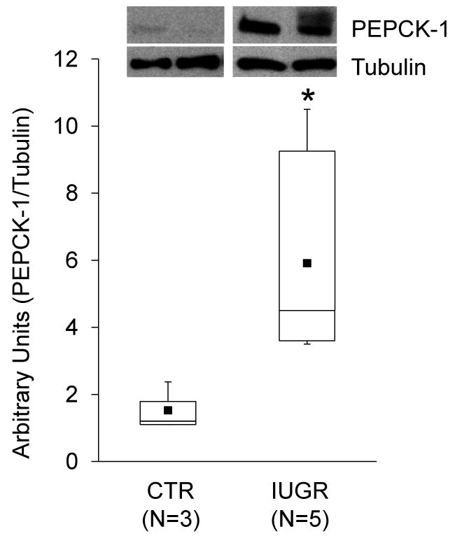
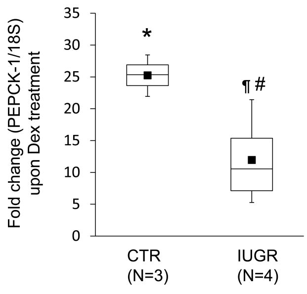
Background: The objective of this study was to develop a cell culture system for fetal baboon hepatocytes and to test the hypotheses that (i) expression of the gluconeogenic enzyme phosphoenolpyruvate carboxykinase-1 (PEPCK-1) is upregulated in hepatocytes isolated from fetuses of nutrient-restricted mothers (MNR) compared with ad libitum-fed controls (CTR), and (ii) glucocorticoids stimulate PEPCK-1 expression.

Methods: Hepatocytes from 0.9G CTR and MNR fetuses were isolated and cultured. PEPCK-1 protein and mRNA levels in hepatocytes were determined by Western blot and quantitative PCR, respectively.

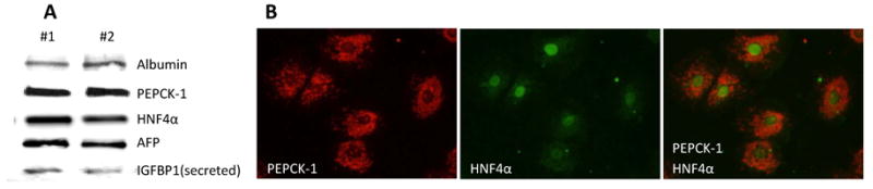
Results: Fetuses of MNR mothers were intrauterine growth restricted (IUGR). Feasibility of culturing 0.9G fetal baboon hepatocytes was demonstrated. PEPCK-1 protein levels were increased in hepatocytes isolated from IUGR fetuses, and PEPCK-1 mRNA expression was stimulated by glucocorticoids in fetal hepatocytes.

Conclusions: Cultured fetal baboon hepatocytes that retain their in vivo phenotype provide powerful in vitro tools to investigate mechanisms that regulate normal and programmed hepatic function.

Keywords: dexamethasone; diabetes non-human primate; liver.


Figures

Figure 1
Figure 1. Expression of hepatocyte markers in 0.9G baboon hepatocytes
A) Western blot analysis demonstrating expression of well-established hepatocyte markers, albumin, PEPCK-1, hepatocyte nuclear factor 4α (HNF4α) and alpha feto protein (AFP), in hepatocytes isolated from 0.9G fetal liver. Presence of secreted insulin-like growth factor binding protein-1 (IGFBP-1) in the media is also observed. B) Immunohistochemistry indicating the presence of cytosolic PEPCK-1 and nuclear HNF4α in hepatocytes isolated from a 0.9G fetus.
Figure 2
Figure 2. PEPCK-1 protein levels are increased in 0.9G baboon hepatocytes isolated from IUGR fetuses
Box-and-whisker plot for CTR (N=3) and IUGR (N=5) are shown. The lower boundary of the box-and-whisker plot corresponds to the 25th percentile, the line within the box to the median, and the upper boundary of the box to the 75th percentile. Mean (■); * (X2=3.76, df=1, P=0.05). Inset shows representative immunoblots from 2 CTR and IUGR fetuses. Tubulin was used as a loading control.
Figure 3
Figure 3. Dexamethasone treatment of hepatocytes from 0.9G CTR and IUGR fetuses increases PEPCK-1 mRNA levels
Box-and-whisker plots depict fold change in PEPCK-1 mRNA levels upon Dex (100 nM) treatment of hepatocytes for 24 h compared to untreated cells. The lower boundary of the box-and-whisker plot corresponds to the 25th percentile, the line within the box to the median, and the upper boundary of the box to the 75th percentile. CTR (N=3, paired); IUGR (N=4, paired); Mean (■); * (X2=4.35, df=1, P<0.04) for Dex-treated vs untreated CTR samples; ¶ (X2=6.05, df=1, P<0.02) for Dex-treated vs untreated IUGR samples; # (X2=4.5, df=1, P<0.04) for CTR vs IUGR Dex-treated samples.

References

    1. Abu Shehab M, Khosravi J, Han VKM, et al. Site-specific IGFBP-1 hyper-phosphorylation in fetal growth restriction: clinical and functional relence. J Proteome Res. 2010;9:1873–1881. - PubMed
    1. Antonow-Schlorke I, Schwab M, Cox LA, et al. Vulnerability of the fetal primate brain to moderate reduction in maternal global nutrient availability. Proc Natl Acad Sci U S A. 2011;108:3011–3016. - PMC - PubMed
    1. Armitage JA, Khan IY, Taylor PD, et al. Developmental programming of the metabolic syndrome by maternal nutritional imbalance: how strong is the evidence from experimental models in mammals? J Physiol. 2004;561:355–377. - PMC - PubMed
    1. Armitage JA, Poston L, Taylor PD. Developmental origins of obesity and the metabolic syndrome: the role of maternal obesity. Front Horm Res. 2008;36:73–84. - PubMed
    1. Armitage JA, Taylor PD, Poston L. Experimental models of developmental programming: consequences of exposure to an energy rich diet during development. J Physiol. 2005;565:3–8. - PMC - PubMed

Publication types

LinkOut - more resources