

Mechanotransduction at focal adhesions: from physiology to cancer development
- PMID: 23601032
- PMCID: PMC3665742
- DOI: 10.1111/jcmm.12045
Mechanotransduction at focal adhesions: from physiology to cancer development
Abstract
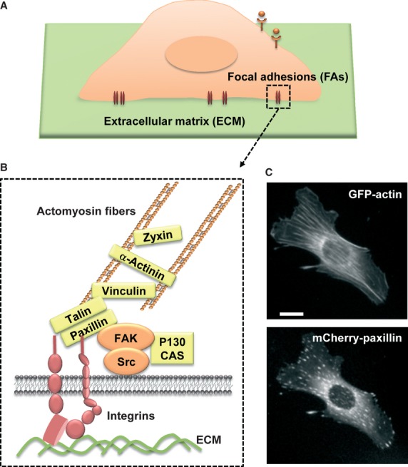
Living cells are continuously exposed to mechanical cues, and can translate these signals into biochemical information (e.g. mechanotransduction). This process is crucial in many normal cellular functions, e.g. cell adhesion, migration, proliferation, and survival, as well as the progression of diseases such as cancer. Focal adhesions are the major sites of interactions between extracellular mechanical environments and intracellular biochemical signalling molecules/cytoskeleton, and hence focal adhesion proteins have been suggested to play important roles in mechanotransduction. Here, we overview the current molecular understanding in mechanotransduction occurring at focal adhesions. We also introduce recent studies on how extracellular matrix and mechanical microenvironments contribute to the development of cancer.
© 2013 The Authors. Published by Foundation for Cellular and Molecular Medicine/Blackwell Publishing Ltd.
Figures
References
-
- Dubash AD, Menold MM, Samson T, et al. Chapter 1. Focal adhesions: new angles on an old structure. Int Rev Cell Mol Biol. 2009;277:1–65. - PubMed
-
- Bershadsky AD, Balaban NQ, Geiger B. Adhesion-dependent cell mechanosensitivity. Annu Rev Cell Dev Biol. 2003;19:677–95. - PubMed
-
- Geiger B, Spatz JP, Bershadsky AD. Environmental sensing through focal adhesions. Nat Rev Mol Cell Biol. 2009;10:21–33. - PubMed
-
- Larsen M, Artym VV, Green JA, et al. The matrix reorganized: extracellular matrix remodeling and integrin signaling. Curr Opin Cell Biol. 2006;18:463–71. - PubMed
Publication types
MeSH terms
Grants and funding
LinkOut - more resources
Full Text Sources
Other Literature Sources
