Comparative Study
. 2013 Aug;28(4):316-30.
doi: 10.1111/omi.12027. Epub 2013 Apr 22.

Sequence divergence in the Treponema denticola FhbB protein and its impact on factor H binding

Affiliations
Comparative Study

Sequence divergence in the Treponema denticola FhbB protein and its impact on factor H binding

D P Miller et al. Mol Oral Microbiol. 2013 Aug.

Abstract

Treponema denticola is an anaerobic spirochete whose abundance in the subgingival crevice correlates with the development and severity of periodontal disease. The ability of T. denticola to survive and thrive in the hostile environment of the periodontal pocket is due, at least in part, to its ability to bind factor H (FH), a negative regulator of the alternative complement pathway. The FH binding protein of T. denticola has been identified as FhbB and its atomic structure has been determined. The interaction of FH with T. denticola is unique in that FH bound to the cell surface is cleaved by the T. denticola protease, dentilisin. It has been postulated that FH cleavage by T. denticola leads to immune dysregulation in periodontal pockets. In this study, we conduct a comparative assessment of the sequence, properties, structure and ligand binding kinetics of the FhbB proteins of strains 33521 and 35405. The biological outcome of the interaction of these strains with FH could differ significantly as 33521 lacks dentilisin activity. The data presented here offer insight into our understanding of the interactions of T. denticola with the host and its potential to influence disease progression.

Keywords: complement; factor H; immune evasion; periodontitis.


Figures

Fig. 1
Fig. 1
FhbB sequence alignment. The DNA sequences of fhbB35405 and fhbB33521 were determined as described in the text and translated. The amino acid sequences were aligned using ClustalW. The leader peptide and structural domains previously determined through X-ray crystallography for the FHbB35405 protein are indicated above the sequences. The FH binding interface of FhbB35405 extends from alpha helix 1 (α1) through alpha helix 2 (α2). Identical residues are indicated by periods and gaps by (−).
Fig. 2
Fig. 2
FhbB variants are antigenically distinct. Recombinant proteins (indicated above each lane) were produced as 6X-His-tagged fusions, purified, subjected to SDS-PAGE and transferred to membranes. The membranes were screened with antiserum as indicated below each panel. Molecular weights are indicated to the left. All methods are detailed in the text.
Fig. 3
Fig. 3
Triton X-114 extraction and phase partitioning of T. denticola demonstrate that FhbB is amphipathic. T. denticola strains 35405 and 33521 were cultivated, harvested, washed, extracted with Triton X114 and phase partitioned as detailed in the text. The resulting fractions (as indicated above each lane) were assessed by SDS-PAGE, transferred to PVDF membranes and screened with anti-FhbB antisera or anti-FlaA antisera. Molecular weight standards are indicated to the left.
Fig. 4
Fig. 4
FhbB is presented on the T. denticola cell surface. Immunofluorescence assays were performed as detailed in the text. Intact (unfixed) or acetone permeabilized (fixed) T. denticola 35405, 33521 and 35405ΔFhbB cells (as indicated to the right of each row of images) were screened with anti-FhbB (α-FhbB) and anti-FlaA (α-FlaA) antiserum. Antibody binding was detected using fluorescently-conjugated antibodies. Cells within each field of view were also visualized by dark-field microscopy.
Fig. 5
Fig. 5
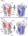
Structural modeling of FhbB. The determined and predicted structures for FhbB35405 (top panel, PDB 3R15) and FhbB33521 (bottom panel) are shown as ribbon diagrams with surface electrostatic charges (red-negative charge; blue-positive charge). The left panel presents a head on view of the FH binding interface. The images in the right panels are rotated 180° around the y-axis. The location of residues that were targeted for site-directed mutagenesis or that are discussed in the text are indicated.
Fig. 6
Fig. 6
Divergent FhbB variants interact with CCP6-7 of FH. R-FhbB35405, FhbB33521, FhbA and BSA were immobilized in the wells of ELISA plates and screened with r-proteins spanning different CCP domains of FH (indicated in the insert). FhbA, which has been demonstrated to bind to CCP19-20, served as a positive control. BSA, which does not bind FH served as a negative control. Binding of the CCP constructs was measured using an ELISA format as detailed in the text.
Fig. 7
Fig. 7
Analysis of the kinetics of the interaction of FH with FhbB35405 and FhbB33521. Binding kinetics were assessed using surface plasmon resonance. R-FhbB proteins were immobilized on a NTA chip and various concentrations of FH were added in the fluid phase. The binding curve obtained using FH at a concentration of 2.5 μM is presented.
Fig. 8
Fig. 8
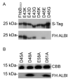
Identification of FhbB residues required for FH binding. Site-directed amino acid substitutions were introduced into FhbB using a mutagenic PCR approach. Recombinant proteins were generated, purified, fractionated by SDS-PAGE and transferred to membranes. Note that the substitution mutants were generated using two different expression vectors and hence the results are presented in two panels. The proteins tested in panel A were generated in pET32 Ek/LIC while those in panel B were generated using pET46 Ek/LIC vector. To verify protein loading, the top blot in panel A was screened with S-Protein as previously described (McDowell et al., 2002). In panel B the protein loading was assessed by staining of a duplicate gel with coomassie brilliant blue G250 (CBB). The membranes were incubated with purified FH and binding detected with FH-specific antisera. As a loading controls, identical blots were screened with HRP-conjugated S-protein to detect the N-terminal expression tag.
Fig. 9
Fig. 9
Strain 33521 lacks dentilisin activity and fails to degrade bound FH. (A) Dentilisin activity of strain 33521 was analyzed by measuring SAAPFNA cleavage. Strain 35405 and 35405-CCE (a dentilisin deficient strain) served as positive and negative controls, respectively. All methods were as detailed in the text. (B) The ability of each strain to cleave FH was assessed through immunoblot analysis of FH that had been incubated with whole cells (as indicated). FH breakdown was monitored using anti-human FH antiserum.
Fig. 10
Fig. 10
Analysis of the sensitivity of T. denticola strains to human serum. Strains 35405 (black), 33521 (dashed lines), and 35405ΔfhbB (white) were incubated in 25% complement-preserved NHS or in HIS for 3 hrs. The percentage of intact cells was determined through dark-field microscopy. The data presented are an average of three individual experiments.

Similar articles

Cited by

References

    1. Bamford CV, Fenno JC, Jenkinson HF, Dymock D. Infect Immun. 2007;75:4364–72. - PMC - PubMed
    1. Beck JD. J Public Health Dent. 1987;47:26–30. - PubMed
    1. Bian XL, Wang HT, Ning Y, Lee SY, Fenno JC. Infect Immun. 2005;73:1252–5. - PMC - PubMed
    1. Boackle RJ. Crit Rev Oral Biol Med. 1991;2:355–67. - PubMed
    1. Boackle RJ, Pruitt KM, Silverman MS, Glymph JL., Jr. J Dent Res. 1978;57:103–10. - PubMed

Publication types

MeSH terms