Review
. 2013 Apr 17;13(4):379-93.
doi: 10.1016/j.chom.2013.03.010.

Virus infections in the nervous system

Affiliations
Review

Virus infections in the nervous system

Orkide O Koyuncu et al. Cell Host Microbe. .

Abstract

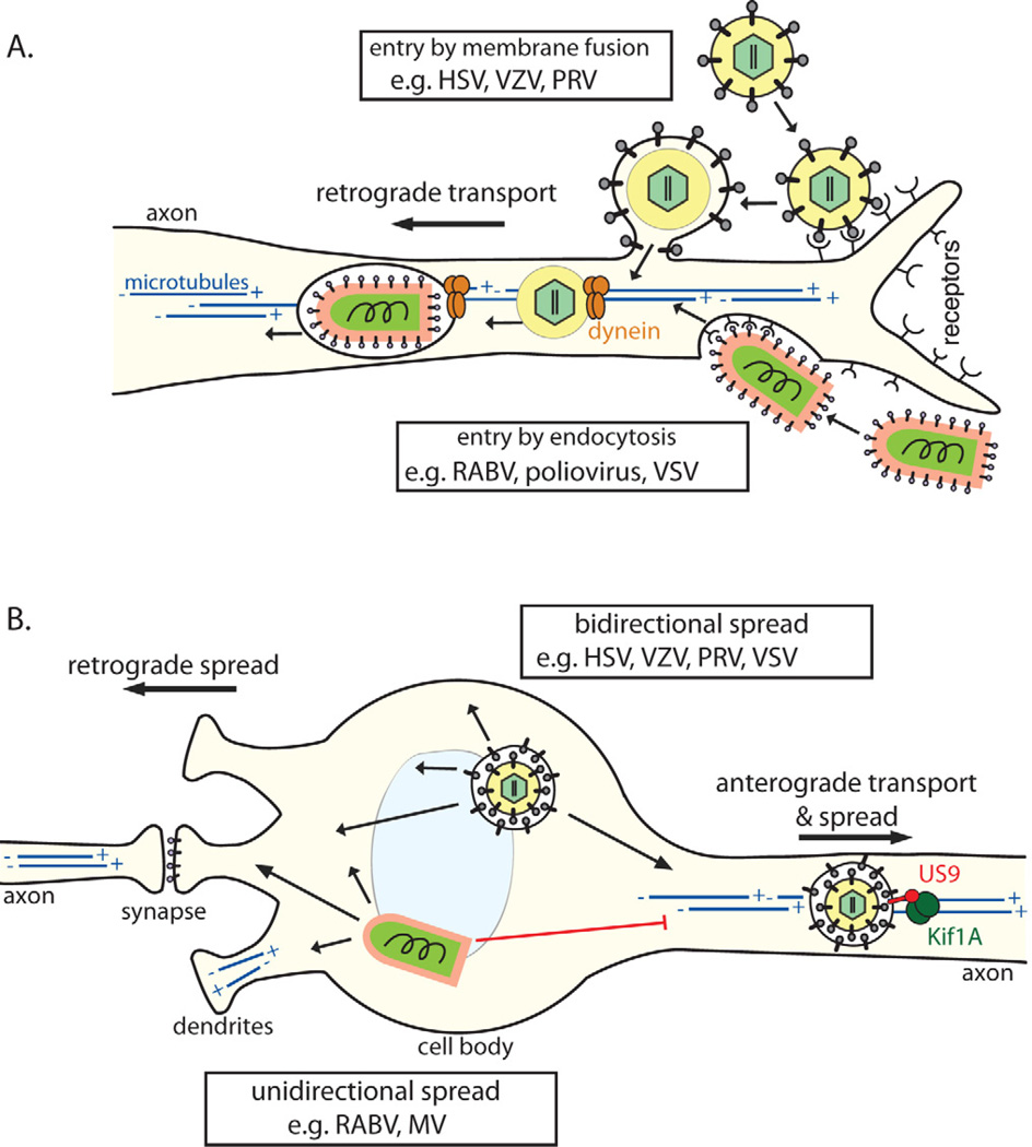
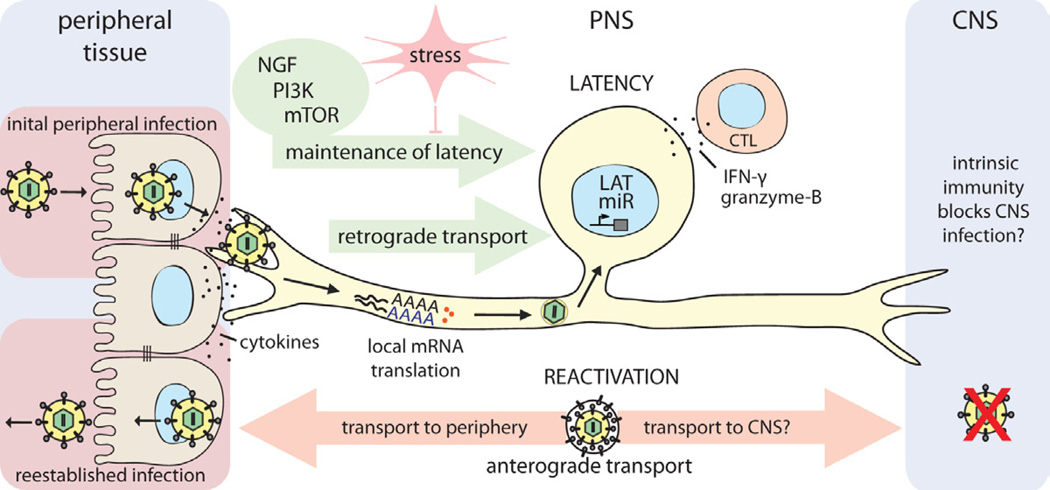
Virus infections usually begin in peripheral tissues and can invade the mammalian nervous system (NS), spreading into the peripheral (PNS) and more rarely the central (CNS) nervous systems. The CNS is protected from most virus infections by effective immune responses and multilayer barriers. However, some viruses enter the NS with high efficiency via the bloodstream or by directly infecting nerves that innervate peripheral tissues, resulting in debilitating direct and immune-mediated pathology. Most viruses in the NS are opportunistic or accidental pathogens, but a few, most notably the alpha herpesviruses and rabies virus, have evolved to enter the NS efficiently and exploit neuronal cell biology. Remarkably, the alpha herpesviruses can establish quiescent infections in the PNS, with rare but often fatal CNS pathology. Here we review how viruses gain access to and spread in the well-protected CNS, with particular emphasis on alpha herpesviruses, which establish and maintain persistent NS infections.


Figures

Figure 1
Figure 1. Immune Control of Viral Infections
Cellular pattern recognition receptors (PRR) recognize viral molecules after attachment and entry. This initial recognition starts a cell autonomous intrinsic defense involving increased synthesis of many antiviral proteins, and several cytokines, including type I interferons (IFN-α/β). If intrinsic defenses fail to stop virus relication, cytokines and infected cell death activate sentinel cells (e.g. dendritic cells, macrophages), which produce copious cytokines and present antigens to trigger T-cell mediated immunity. The innate immune response, which is activated quickly and robustly, also involves the actions of the complement system, natural killer cells (NK), neutrophils and other granulocytes. Over-reaction of this response causes immunopathology termed a “cytokine storm”. The adaptive, acquired immune response is slow, systemic, pathogen-specific, and leads to the induction of immunological memory. The cell-mediated “Th1” response involves the action of CD4+ T-helper cells and CD8+ Cytotoxic T-cells (CTL). CTLs are important to kill infected cells and to produce type II interferon (IFN-γ) and tumor necrosis factor (TNF-α).The “Th2” humoral response involves CD4+ T-helper cells and antibody-producing B-cells. Most of the time, the clinical symptoms of virus infection (e.g. fever, pain, tissue damage) are caused by the inflammatory action of the innate and adaptive immune systems. Due to their mostly irreplacable nature, nervous system tissues rely predominantly on the intrinsic and innate immune responses, and avoid the extensive inflammation and cytotoxic effects of the adaptive immune response.
Figure 2
Figure 2. Virus entry routes into the CNS
(A) Alpha herpesviruses (e.g. HSV-1, VZV, PRV) infect pseudounipolar sensory neurons of PNS ganglia. CNS spread is rare and requires anterograde axonal transport of progeny virions toward the spinal cord. (B) RABV and poliovirus spread via neuromuscular junctions (NMJ) from muscles into somatic motor neurons in the spinal cord. (C) Several viruses may infect receptor neurons in the nasal olfactory epithelium. Spread to the CNS requires anterograde axonal transport along the olfactory nerve into the brain. (D) Infiltration through the blood-brain barrier (BBB). The BBB is composed of brain microvascular endothelium cells (BMVEC) with specialized tight junctions, surrounding basement membrane, pericytes, astrocytes, and neurons. Infected leukocytes can traverse this barrier carrying virus into the brain parenchyma. (E) Alternatively, virus particles in the bloodstream can infect BMVECs, compromising the BBB.
Figure 3
Figure 3. Cell biology of virus entry, transport, and spread in the NS
(A) Virons enter axons either via (1) direct fusion with the axonal membrane after receptor attachment (e.g. alpha herpesviruses) or by (2) endocytosis (e.g. RABV; most neurotropic viruses). All particles entering the axon cytoplasm require (−) end-directed dynein motors for long distance retrograde transport on microtubules toward the cell body. Microtubules in axons are uniformly oriented with (+) ends facing the axon terminus and the (−) ends in the cell body. In the cell body and dendrites, microtubules have mixed polarity. (B) Post-replication trafficking and spread of progeny virions. Progeny alpha herpesvirus virions in transport vesicles can be transported anterograde in axons, dependent on the viral protein Us9 associating with the microtubule motor kinesin-3/Kif1A. Egress from axons allows anterograde virus spread from pre-synaptic to post-synaptic neurons. Virions in transport vesicles can also traffic to and egress from the somatodendritic compartment, allowing retrograde spread from post-synaptic to presynaptic neurons. Accordingly, herpesviruses can spread bidirectionally (1) in neuronal circuits. In contrast, enveloped RNA virions (e.g. RABV, MV) acquire their membranes by budding though the plasma membrane during egress. For these viruses, the location of envelopment/egress, and directionality of spread, may depend on the transport and intracellular localization of viral glycoproteins. These viruses tend to spread unidirectionally (2), only from post-synaptic to pre-synaptic neurons.
Figure 4
Figure 4. Establishment and control of alpha herpesvirus latency in PNS neurons
HSV-1 enters the host organism through mucosal/epithelial surfaces. Initial replication in epithelial cells results both in cytokine secretion and viral spread. Progeny virions enter sensory nerve terminals and undergo long-distance retrograde transport to the neuronal cell body. Localized intracellular signaling and protein translation in distal axons induced by cytokines and viral proteins during entry supports retrograde particle transport and also may influence the establishment of latency in neuronal cell bodies. Once in the nucleus, viral latency genes are activated (e.g. LAT, miRs), and the HSV-1 genome is repressed. CD8+ CTLs secreting IFN-γ and granzyme-B, and continuous retrograde NGF-PI3K-mTOR signaling from distal axons contribute to maintaining this latent state of infection. If this homeostasis is disrupted by environmental or nutritional stress, the HSV-1 genome reactivates lytic gene expression, leading to virus replication. Newly replicated virions traffic back by anterograde axonal transport mechanisms, to reestablish infection at epithelial tissues. It is still not understood why CNS invasion is rare after reactivation from latency, even though both axonal projections of the pseudounipolar sensory neuron are accessible to HSV-1 virion transport. If there is no difference in transport, it may be that HSV-1 does invade the CNS, but is strongly suppressed or silenced by an effective intrinsic immune response in CNS neurons of healthy individuals.

References

    1. Teijaro JR, et al. Endothelial cells are central orchestrators of cytokine amplification during influenza virus infection. Cell. 146(6):980–991. - PMC - PubMed
    1. Griffin DE, Metcalf T. Clearance of virus infection from the CNS. Curr Opin Virol. 2011;1(3):216–221. - PMC - PubMed
    1. Tirabassi RS, et al. Molecular mechanisms of neurotropic herpesvirus invasion and spread in the CNS. Neurosci Biobehav Rev. 1998;22(6):709–720. - PubMed
    1. Smith G. Herpesvirus transport to the nervous system and back again. Annu Rev Microbiol. 2012;66:153–176. - PMC - PubMed
    1. Camarena V, et al. Nature and duration of growth factor signaling through receptor tyrosine kinases regulates HSV-1 latency in neurons. Cell Host Microbe. 2010;8(4):320–330. - PMC - PubMed

Publication types

MeSH terms