. 2013 Apr 17;13(4):417-28.
doi: 10.1016/j.chom.2013.03.001.

Disease-enhancing antibodies improve the efficacy of bacterial toxin-neutralizing antibodies

Affiliations

Disease-enhancing antibodies improve the efficacy of bacterial toxin-neutralizing antibodies

Siu-Kei Chow et al. Cell Host Microbe. .

Abstract

During infection, humoral immunity produces a polyclonal response with various immunoglobulins recognizing different epitopes within the microbe or toxin. Despite this diverse response, the biological activity of an antibody (Ab) is usually assessed by the action of a monoclonal population. We demonstrate that a combination of monoclonal antibodies (mAbs) that are individually disease enhancing or neutralizing to Bacillus anthracis protective antigen (PA), a component of anthrax toxin, results in significantly augmented protection against the toxin. This boosted protection is Fc gamma receptor (FcγR) dependent and involves the formation of stoichiometrically defined mAb-PA complexes that requires immunoglobulin bivalence and simultaneous interaction between PA and the two mAbs. The formation of these mAb-PA complexes inhibits PA oligomerization, resulting in protection. These data suggest that functional assessments of single Abs may inaccurately predict how the same Abs will operate in polyclonal preparations and imply that potentially therapeutic mAbs may be overlooked in single Ab screens.


Figures

Figure 1
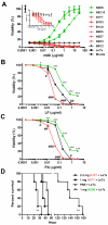
Figure 1. see also Figure S1 and Table S1. Protective and disease-enhancing activities of PA-specific mAbs
(A) The protective efficacy of individual mAbs was measured by MTT assay with J774.16 cells. The viabilities of cells treated with media or LeTx only were used as references to determine mAbs to be protective (green), indifferent (black), or enhancing (red). The inset is the magnification of the region showing only indifferent (black) and enhancing (red) mAbs. (B–C) The effect of protective mAb N2D6 (green) and enhancing mAb N1F7 (red) on LeTx toxicity studied by MTT assay using J774.16 macrophage-like cells, with (B) PA concentration being constant (0.5 μg/ml) and the one of LF titrated, and (C) LF concentration being constant (0.5 μg/ml) and the one of PA titrated vice versa. mAb concentration = 5 μg/ml. error bars, mean ± SD; ***P < 0.0005, **P < 0.005, *P < 0.05 protective mAb versus no mAb; ###P <0.0005, ##P < 0.005, #P < 0.05 enhancing mAb versus no mAb, all by t test. (D) Dose dependent survival of BALB/c mice challenged with LeTx (35 μg of PA and LF each) 4 h after pre-injection of protective mAb N2D6 or enhancing mAb N1F7. PBS control was used as reference. Data shown are representative of three independent experiments. Means and SD of triplicates are shown from one representative experiment out of three (A–C). In vivo studies are representative of two independent experiments with n = 5 mice per group; **P < 0.005, *P < 0.05 log-rank test.
Figure 2
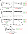
Figure 2. Combination of protective and enhancing mAbs to PA boosts protection against LeTx
The protective efficacy of enhancing mAbs (red) in the presence of protective mAbs (green), (A) N2D6, (B) N5C10, (C) 19D9, respectively, measured by MTT assay, with the concentration of protective mAbs being constant (1 μg/ml). (D) The protective efficacy of protective mAbs (green) and indifferent mAb (black) was determined by MTT assay in the absence (dotted line) and presence (solid line) of enhancing mAb, N1F7 (red), with the concentration of N1F7 being constant (0.1 μg/ml). mAb 19D9 was described by Abboud et al (2010). error bars, mean ± SD; ***P < 0.0005 absence of N1F7 versus corresponding presence of N1F7 counterparts, by t test. (E) LDH measurement mirrored results from the MTT assay to show significant protection against LeTx by combining protective and enhancing mAbs. (top panel) MTT assay showed the protective efficacy against LeTx of protective mAb (green), enhancing mAb (red), and their mixture (black). (bottom panel) LDH measurement showed the same protection pattern as MTT assay. (F) Percentage survival of BALB/c mice challenged with LeTx (35 μg of PA and LF each) 4 h after pre-injection of PBS, individual mAb (N2D6 as protective, N1F7 as enhancing), or mAb mixture (1 mg mAb each per mouse). The irrelevant mAb was 18B7, an IgG1 murine mAb that did not bind to PA or LF (Mukherjee et al., 1992). In vivo studies are representative of two independent experiments with n = 5 mice per group; **P < 0.005, *P < 0.05 log-rank test. Data shown are representative of two independent experiments. Means and SD of triplicates are shown from one representative experiment out of two (A–E).
Figure 3
Figure 3. Epitope competition of the protective and enhancing mAbs results in no synergistic protection
Competition binding assay for PA between protective mAbs (green) and enhancing mAbs (red) as determined using inhibition ELISA. The concentration of protective mAbs (A) N2D6, (B) N5C10, (C) 19D9, respectively, was kept constant (2 μg/ml), and those of enhancing mAbs were titrated, and vice versa. (D) The protective efficacy of whole IgG mAbs compared with their Fab fragments by MTT assay. The concentration of N2D6 whole IgG and Fab is 6.67 nM. Data shown are representative of three independent experiments. Means and SD of triplicates are shown from one representative experiment out of three (A–D).
Figure 4
Figure 4. see also Figure S2. Immunoglobulin bivalence and FcγR engagement contribute to protective synergy
(A) The function of Fc region in Ab-mediated protection was measured by comparing LeTx cytotoxicity on bone marrow macrophages (BMMs) derived from FcRγ chain/FcγRII double knockout (FcRγ−/−/RIIB−/−) and wild type (WT) mice using MTT assay. error bars, mean ± SD; **P < 0.005, *P < 0.05 WT BMM N2D6 with N1F7whole IgG versus with N1F7Fab;&&P < 0.005,&P < 0.05 DKO BMM N2D6 with N1F7whole IgG versus with N1F7Fab, all by t test. N2D6 concentration was kept constant (6.67 nM). (B) The protective efficacy of whole IgG mAbs was compared with their F(ab')2 fragments by MTT assay. N2D6 concentration was kept constant (6.67 nM). (C) Percentage survival of BALB/c mice challenged with LeTx 4 h after pre-injection of PBS, mAb F(ab')2, mAb whole IgG, or mAb mixture (1 mg whole IgG or 0.667 mg F(ab')2 each). Representative of two independent experiments with n = 5 mice per group; **P < 0.005, log-rank test. (D) Percentage survival of wild type C57BL/6 and FcRγ−/−/RIIB−/− mice challenged with LeTx pre-injected with 1:1 mixture of N2D6 and N1F7 (1 mg each). P = 0.0005, log-rank test. Data shown are representative of three independent experiments. (E–G) Switching of Fc region on enhancing mAbs did not affect protective outcome when mixed with protective mAb. IgG1 enhancing mAbs, (E) N5D7 and (F) N4H3, were class switched to IgG2a and IgG2b, respectively. MTT assay indicated no significant change of LeTx enhancing activities for the switched mAbs. (G) Different classes of N5D7 and N4H3 were mixed with protective mAb, N2D6 (1 μg/ml), and the combinational protective outcome was determined by MTT assay. Data shown are representative of three independent experiments. Means and SD of triplicates are shown from one representative experiment out of three (A–B, E–G).
Figure 5
Figure 5. Molar ratio of protective and enhancing mAbs determine mAb-PA complex formation
Protection from combining protective and enhancing mAbs peaked at optimum molar ratio of the two. (A) The protection from N2D6 with minimal amount of N1F7 (<0.2 ng/ml) was set as baseline protection, which increased with the concentration of N2D6. (B) To reveal the synergistic protection given solely by the addition of N1F7 to the system, the baseline protection of individual N2D6 concentration was subtracted from the corresponding plot of mixing N2D6 and N1F7. The two-dimensional plots illustrated the results after normalization. (C) Three-dimensional plot of net protection given by the mixing of protective and enhancing mAbs with respect to protective mAb alone. The concentration of PA and LF used in this experiment was 0.6 μg/ml each. Data shown are representative of two independent experiments. (A–C). (D) Alexa-Fluor−568, −405, and−488 conjugated protective mAb, enhancing mAb, and PA mixed in molar ratio 1:0.001:1 (X), 1:1:1 (Y), or 1:20:1 (Z), and were added to J774.16 macrophages to allow cell-based mAb-PA interaction but not phagocytosis by the cells. The cells were imaged by epifluorescence microscopy. n > 150 cells, ***P < 0.0005 and **P < 0.005, t test. Scale bar = 30 μm (E) J774.16 macrophage-like cells were incubated with Alexa-Fluor conjugated mAbs and PA as described in (D) followed by phagocytosis of the mAb-PA complexes by macrophages. The uptake was measured by flow cytometry, and presented as total cell-associated fluorescence. Data shown are representative of three independent experiments. Means and SD of triplicates are shown from one representative experiment out of three (D and E).
Figure 6
Figure 6. see also Figure S3. Experimental mAb-PA complex formation with discrete molecular weights concurs with computational model
(A) Individual mAbs or their mixture were incubated with PA and subjected to native PAGE followed by Coomassie Blue staining, which aimed to study the mAb-PA complex formation with their natural conformations. The irrelevant mAb was 18B7 (Mukherjee et al., 1992). (B) Individual mAbs or their mixture were incubated with PA, and their interactions were stabilized by crosslinker BS3. The reaction mixture was subjected to SDS-PAGE followed by Western blotting. A PA-specific mAb, 10F4 (IgG1), that targeted PA63 region of the PA molecule was used to probe PA and its complexes with mAbs. (C) Schematic diagram showing the components of mAb-PA complexes with distinct molecular weight and comparison of gel band optical density to rule-based model. A Kolmogorov-Smirnov test adapted for discrete distributions did not reject the null hypothesis that the empirical and model distributions represent samples from the same underlying form. Error bars show sample SD. Data shown are representative of three independent experiments. SD of triplicates is shown from one representative experiment out of three (C).
Figure 7
Figure 7. see also Table S1. mAb-PA complex formation abrogates PA oligomerization
(A) PA pre-incubated with individual mAbs, or mAb mixture was added to J774.16 macrophages and incubated for 45 min to allow furin cleavage, oligomer formation and uptake by the cells. Whole cell lysate was subjected to SDS-PAGE and Western blotting probed with PA-specific mAb 10F4 (IgG1) that recognized PA83, PA63, and the PA oligomers. Data shown are representative of three independent experiments. (B) Protective efficacy of enhancing mAbs (red) in the presence of protective mAbs 10F4 (green) measured by MTT, with the concentration of 10F4 being constant as 1 μg/ml. 10F4 binds to a distant epitope from the PA20 region of PA83, as described by Rivera at al. (2006). Data shown are representative of two independent experiments. SD of triplicates are shown from one representative experiment out of two. (C) Proposed mechanism for synergistic protection against LeTx involving the combination of protective and enhancing mAbs.

Similar articles

Cited by

References

    1. Abboud N, Chow SK, Saylor C, Janda A, Ravetch JV, Scharff MD, Casadevall A. A requirement for FcgammaR in antibody-mediated bacterial toxin neutralization. J Exp Med. 2010;207:2395–2405. - PMC - PubMed
    1. Abboud N, De Jesus M, Nakouzi A, Cordero RJ, Pujato M, Fiser A, Rivera J, Casadevall A. Identification of linear epitopes in Bacillus anthracis protective antigen bound by neutralizing antibodies. J Biol Chem. 2009;284:25077–25086. - PMC - PubMed
    1. Ablowitz The theory of emergence. Phil Sci. 1939;6:1–16.
    1. Aderem A, Underhill DM. Mechanisms of phagocytosis in macrophages. Annu Rev Immunol. 1999;17:593–623. - PubMed
    1. Anderson P, Caligiuri M, O'Brien C, Manley T, Ritz J, Schlossman SF. Fc gamma receptor type III (CD16) is included in the zeta NK receptor complex expressed by human natural killer cells. Proc Natl Acad Sci U S A. 1990;87:2274–2278. - PMC - PubMed

Publication types

LinkOut - more resources