

The effect of computed tomographic scanner parameters and 3-dimensional volume rendering techniques on the accuracy of linear, angular, and volumetric measurements of the mandible
- PMID: 23601224
- PMCID: PMC3633147
- DOI: 10.1016/j.oooo.2013.02.008
The effect of computed tomographic scanner parameters and 3-dimensional volume rendering techniques on the accuracy of linear, angular, and volumetric measurements of the mandible
Abstract
Objectives: This study investigates the effect of scanning parameters on the accuracy of measurements from three-dimensional (3D), multi-detector computed tomography (MDCT) mandible renderings. A broader range of acceptable parameters can increase the availability of computed tomographic (CT) studies for retrospective analysis.
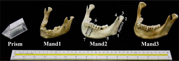
Study design: Three human mandibles and a phantom object were scanned using 18 combinations of slice thickness, field of view (FOV), and reconstruction algorithm and 3 different threshold-based segmentations. Measurements of 3D computed tomography (3DCT) models and specimens were compared.
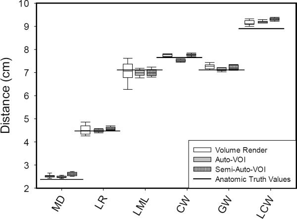
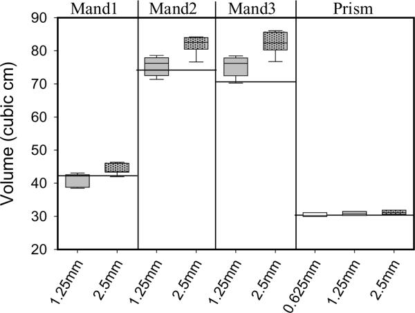
Results: Linear and angular measurements were accurate, irrespective of scanner parameters or rendering technique. Volume measurements were accurate with a slice thickness of 1.25 mm, but not 2.5 mm. Surface area measurements were consistently inflated.
Conclusions: Linear, angular, and volumetric measurements of mandible 3D MDCT models can be confidently obtained from a range of parameters and rendering techniques. Slice thickness is the primary factor affecting volume measurements. These findings should also apply to 3D rendering using cone-beam CT (CBCT).
Copyright © 2013 Elsevier Inc. All rights reserved.
Figures
References
-
- Nkenke E, Zachow S, Benz M, Maier T, Veit K, Kramer M, et al. Fusion of computed tomography data and optical 3D images of the dentition for streak artefact correction in the simulation of orthognathic surgery. Dentomaxillofac Radiol. 2004;33:226–32. - PubMed
-
- Pohlenz P, Blessmann M, Blake F, Gbara A, Schmelzle R, Heiland M. Major mandibular surgical procedures as an indication for intraoperative imaging. J Oral Maxillofac Surg. 2008;66(2):324–9. - PubMed
-
- Hashimoto K, Arai Y, Iwai K, Araki M, Kawashima S, Terakado M. A comparison of a new limited cone beam computed tomography machine for dental use with a multidetector row helical CT machine. Oral Surg Oral Med Oral Path Oral Radiol Endod. 2003;95(3):371–377. - PubMed
-
- Mozzo P, Procacci C, Tacconi A, Martini PT, Andreis IA. A new volumetric CT machine for dental imaging based on the cone-beam technique: preliminary results. Eur Radiol. 1998;8(9):1558–1564. - PubMed
Publication types
MeSH terms
Substances
Grants and funding
LinkOut - more resources
Full Text Sources
Other Literature Sources
