

Constant or fluctuating hyperglycemias increases cytomembrane stiffness of human umbilical vein endothelial cells in culture: roles of cytoskeletal rearrangement and nitric oxide synthesis
- PMID: 23601245
- PMCID: PMC3651398
- DOI: 10.1186/1471-2121-14-22
Constant or fluctuating hyperglycemias increases cytomembrane stiffness of human umbilical vein endothelial cells in culture: roles of cytoskeletal rearrangement and nitric oxide synthesis
Abstract
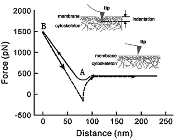
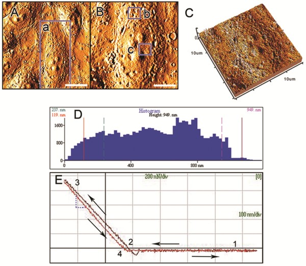
Background: Previous studies have implicated continuous or intermittent hyperglycemia in altered endothelium-derived nitric oxide (NO) synthesis. NO can regulate both the F-actin cytoskeleton and endothelial cell membrane stiffness. Atomic force microscopy (AFM) is a powerful tool that can be used to study plasma membrane deformability at the single cell level. As membrane stiffness is partially dependent on filamentous F-actin, the interdependence of these parameters can be studied through the combined approaches of AFM and laser scanning confocal microscopy (LSCM). In the present study, we evaluated the effects of constant or fluctuating hyperglycemia on endothelial-derived NO synthesis, the cytoskeletal contribution and endothelial cell membrane stiffness.
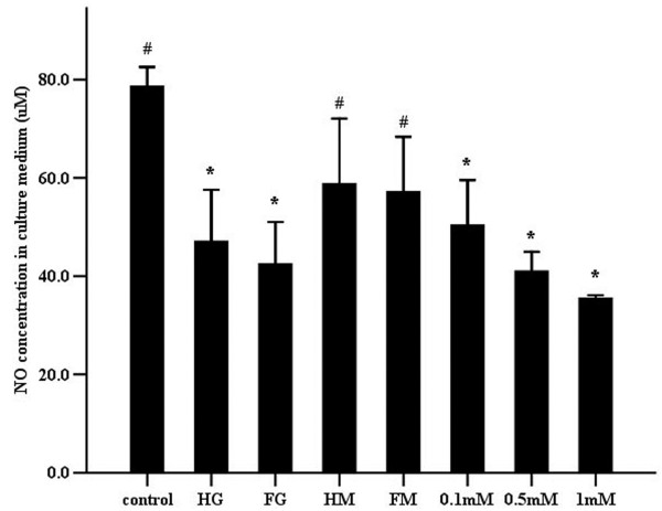
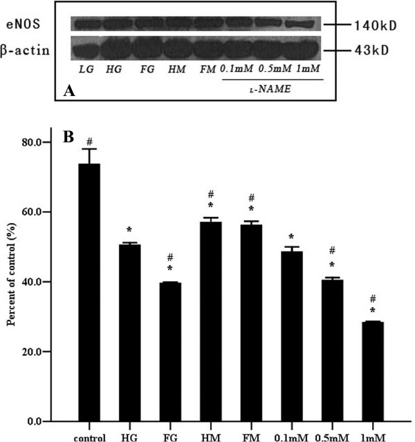
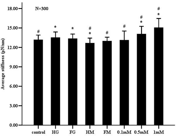
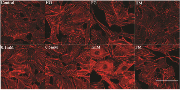
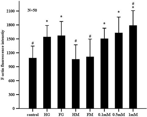
Results: Compared to control cells cultured in low glucose (5 mM), constant (25 mM) or fluctuating (25/5 mM) high glucose significantly decreased NO release along with stiffening of endothelial cell membranes and F-actin rearrangement. The non-selective nitric oxide synthase (NOS) inhibitor, N(G)-nitro-(L)-arginine methyl ester ((L)-NAME) exerted similar effects on endothelial cells. Increasing concentrations of (L)-NAME (from 0.1 to 1 mM) exacerbated these effects in a concentration-dependent manner.
Conclusions: Result from the present study suggest that stiffening endothelial cell membranes are associated with decreased NO synthesis, which was established through the F-actin cytoskeletal redistribution. The precise mechanisms of hyperglycemia-induced endothelial dysfunction require further investigation.
Figures
Similar articles
-
Mechanisms underlying beta2-adrenoceptor-mediated nitric oxide generation by human umbilical vein endothelial cells.J Physiol. 2006 Oct 15;576(Pt 2):585-94. doi: 10.1113/jphysiol.2006.115998. Epub 2006 Jul 27. J Physiol. 2006. PMID: 16873402 Free PMC article.
-
Nitric oxide and cGMP regulate endothelial permeability and F-actin distribution in hydrogen peroxide-treated endothelial cells.Exp Cell Res. 1997 Aug 25;235(1):238-44. doi: 10.1006/excr.1997.3675. Exp Cell Res. 1997. PMID: 9281373
-
Inhibition of nitric oxide synthase (NOS) conversion of L-arginine to nitric oxide (NO) decreases low density mononuclear cell (LD MNC) trans-endothelial migration and cytokine output.J Surg Res. 2003 Sep;114(1):100-6. doi: 10.1016/s0022-4804(03)00310-x. J Surg Res. 2003. PMID: 13678705
-
A role for insulin on L-arginine transport in fetal endothelial dysfunction in hyperglycaemia.Curr Vasc Pharmacol. 2009 Oct;7(4):467-74. doi: 10.2174/157016109789043919. Curr Vasc Pharmacol. 2009. PMID: 19485892 Review.
-
Old and New Biomarkers Associated with Endothelial Dysfunction in Chronic Hyperglycemia.Oxid Med Cell Longev. 2021 Dec 27;2021:7887426. doi: 10.1155/2021/7887426. eCollection 2021. Oxid Med Cell Longev. 2021. PMID: 34987703 Free PMC article. Review.
Cited by
-
High glucose induces and activates Toll-like receptor 4 in endothelial cells of diabetic retinopathy.Diabetol Metab Syndr. 2015 Oct 13;7:89. doi: 10.1186/s13098-015-0086-4. eCollection 2015. Diabetol Metab Syndr. 2015. PMID: 26468333 Free PMC article.
-
Aberrant phenotype in human endothelial cells of diabetic origin: implications for saphenous vein graft failure?J Diabetes Res. 2015;2015:409432. doi: 10.1155/2015/409432. Epub 2015 Apr 8. J Diabetes Res. 2015. PMID: 25950006 Free PMC article.
-
Advanced Glycation End Products Effects on Adipocyte Niche Stiffness and Cell Signaling.Int J Mol Sci. 2023 Jan 23;24(3):2261. doi: 10.3390/ijms24032261. Int J Mol Sci. 2023. PMID: 36768583 Free PMC article.
-
Hyperglycemia-Induced Changes in Hyaluronan Contribute to Impaired Skin Wound Healing in Diabetes: Review and Perspective.Int J Cell Biol. 2015;2015:701738. doi: 10.1155/2015/701738. Epub 2015 Sep 10. Int J Cell Biol. 2015. PMID: 26448756 Free PMC article. Review.
-
Reduced cofilin activity as a mechanism contributing to endothelial cell stiffening in type 2 diabetes.Am J Physiol Heart Circ Physiol. 2025 Jan 1;328(1):H84-H92. doi: 10.1152/ajpheart.00667.2024. Epub 2024 Nov 29. Am J Physiol Heart Circ Physiol. 2025. PMID: 39611817 Free PMC article.
References
-
- UK Prospective Diabetes Study (UKPDS) Group. Intensive blood-glucose control with sulphonylureas or insulin compared with conventional treatment and risk of complications in patients with type 2 diabetes (UKPDS 33) Lancet. 1998;352:837–853. - PubMed
-
- Piconi L, Quagliaro L, Da RR, Assaloni R, Giugliano D, Esposito K, Szabo C, Ceriello A. Intermittent high glucose enhances ICAM-1, VICAM-1, E-selectin and interleukin-6 expression in human umbilical enclothelial cells in culture: the role of poly(ADP-ribose) polymerase. J Thromb Haemost. 2004;2:1453–1459. doi: 10.1111/j.1538-7836.2004.00835.x. - DOI - PubMed
-
- Quagliaro L, Piconi L, Assaloni R, Da RR, Maier A, Zuodar G, Ceriello A. Intermittent high glucose enhances ICAM-1, VCAM-1 and E-selectin expression in human umbilical vein endothelial cells in culture: the distinct role of protein kinase C and mitochondrial superoxide production. Atherosclerosis. 2005;183:259–267. doi: 10.1016/j.atherosclerosis.2005.03.015. - DOI - PubMed
-
- Pricci F, Leto G, Amadio L, Iacobini C, Cordone S, Catalano S, Zicari A, Sorcini M, Di MU, Pugliese G. Oxidative stress in diabetes-induced endothelial dysfunction involvement of nitric oxide and protein kinase C. Free Radic Biol Med. 2003;35:683–694. doi: 10.1016/S0891-5849(03)00401-5. - DOI - PubMed
Publication types
MeSH terms
Substances
LinkOut - more resources
Full Text Sources
Other Literature Sources
Medical
Research Materials
Miscellaneous
