

Cell cycle-dependent binding modes of the ran exchange factor RCC1 to chromatin
- PMID: 23601311
- PMCID: PMC3627872
- DOI: 10.1016/j.bpj.2013.03.024
Cell cycle-dependent binding modes of the ran exchange factor RCC1 to chromatin
Abstract
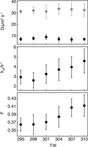
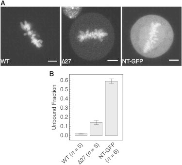
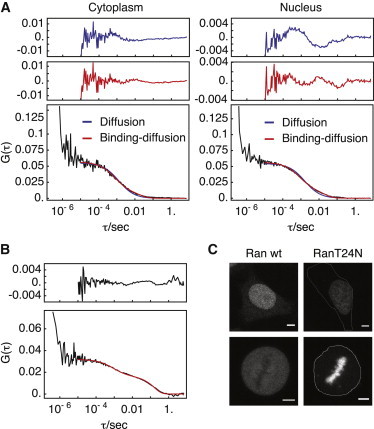
The formation of an activity gradient of the small G-protein Ran around chromatin depends on the differential partitioning of the opposing enzyme activities of the Ran guanine nucleotide exchange factor RCC1 that resides on chromatin, and the cytoplasmic Ran GTPase activating protein RanGAP. We studied the time-dependent interaction kinetics between RCC1 and chromatin and the mobility of the Ran-RCC1 complex in living cells by fluorescence correlation spectroscopy to investigate whether binding of RCC1 to chromatin regulates the exchange activity of RCC1, and whether the stability of the RCC1-chromatin interaction is regulated during the cell cycle. We found that RCC1 mobility is dominated by two states: a highly mobile state that is trapped within chromatin, and a transiently immobilized state that is stabilized during mitosis. We show that only the immobilized state of RCC1 interacts with Ran and conclude that its guanine nucleotide exchange activity is restricted to specific sites on chromatin.
Copyright © 2013 Biophysical Society. Published by Elsevier Inc. All rights reserved.
Figures


Similar articles
-
RanBP1 controls the Ran pathway in mammalian cells through regulation of mitotic RCC1 dynamics.Cell Cycle. 2020 Aug;19(15):1899-1916. doi: 10.1080/15384101.2020.1782036. Epub 2020 Jun 28. Cell Cycle. 2020. PMID: 32594833 Free PMC article.
-
The methylated N-terminal tail of RCC1 is required for stabilisation of its interaction with chromatin by Ran in live cells.BMC Cell Biol. 2010 Jun 21;11:43. doi: 10.1186/1471-2121-11-43. BMC Cell Biol. 2010. PMID: 20565941 Free PMC article.
-
Regulation of chromatin binding by a conformational switch in the tail of the Ran exchange factor RCC1.J Cell Biol. 2008 Sep 8;182(5):827-36. doi: 10.1083/jcb.200803110. Epub 2008 Sep 1. J Cell Biol. 2008. PMID: 18762580 Free PMC article.
-
Premature chromatin condensation caused by loss of RCC1.Prog Cell Cycle Res. 2000;4:145-56. doi: 10.1007/978-1-4615-4253-7_13. Prog Cell Cycle Res. 2000. PMID: 10740822 Review.
-
RCC1 in the Ran pathway.J Biochem. 1996 Aug;120(2):207-14. doi: 10.1093/oxfordjournals.jbchem.a021400. J Biochem. 1996. PMID: 8889801 Review.
Cited by
-
Axon-soma communication in neuronal injury.Nat Rev Neurosci. 2014 Jan;15(1):32-42. doi: 10.1038/nrn3609. Epub 2013 Dec 11. Nat Rev Neurosci. 2014. PMID: 24326686 Review.
-
EBNA1: Oncogenic Activity, Immune Evasion and Biochemical Functions Provide Targets for Novel Therapeutic Strategies against Epstein-Barr Virus- Associated Cancers.Cancers (Basel). 2018 Apr 6;10(4):109. doi: 10.3390/cancers10040109. Cancers (Basel). 2018. PMID: 29642420 Free PMC article. Review.
-
Phosphorylation of RCC1 on Serine 11 Facilitates G1/S Transition in HPV E7-Expressing Cells.Biomolecules. 2021 Jul 6;11(7):995. doi: 10.3390/biom11070995. Biomolecules. 2021. PMID: 34356619 Free PMC article.
-
Contractility kits promote assembly of the mechanoresponsive cytoskeletal network.J Cell Sci. 2019 Jan 16;132(2):jcs226704. doi: 10.1242/jcs.226704. J Cell Sci. 2019. PMID: 30559246 Free PMC article.
-
Nucleosome functions in spindle assembly and nuclear envelope formation.Bioessays. 2015 Oct;37(10):1074-85. doi: 10.1002/bies.201500045. Epub 2015 Jul 29. Bioessays. 2015. PMID: 26222742 Free PMC article. Review.
References
-
- Clarke P.R., Zhang C. Spatial and temporal coordination of mitosis by Ran GTPase. Nat. Rev. Mol. Cell Biol. 2008;9:464–477. - PubMed
-
- Kaláb P., Pralle A., Weis K. Analysis of a RanGTP-regulated gradient in mitotic somatic cells. Nature. 2006;440:697–701. - PubMed
-
- Caudron M., Bunt G., Karsenti E. Spatial coordination of spindle assembly by chromosome-mediated signaling gradients. Science. 2005;309:1373–1376. - PubMed
-
- Nemergut M.E., Mizzen C.A., Macara I.G. Chromatin docking and exchange activity enhancement of RCC1 by histones H2A and H2B. Science. 2001;292:1540–1543. - PubMed
-
- Trieselmann N., Wilde A. Ran localizes around the microtubule spindle in vivo during mitosis in Drosophila embryos. Curr. Biol. 2002;12:1124–1129. - PubMed
Publication types
MeSH terms
Substances
LinkOut - more resources
Full Text Sources
Other Literature Sources
Molecular Biology Databases
Miscellaneous
