

Cooperative heparin-mediated oligomerization of fibroblast growth factor-1 (FGF1) precedes recruitment of FGFR2 to ternary complexes
- PMID: 23601319
- PMCID: PMC3628569
- DOI: 10.1016/j.bpj.2013.02.051
Cooperative heparin-mediated oligomerization of fibroblast growth factor-1 (FGF1) precedes recruitment of FGFR2 to ternary complexes
Abstract
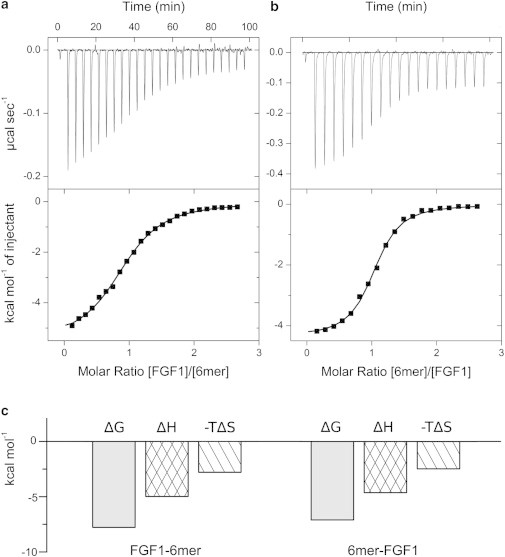
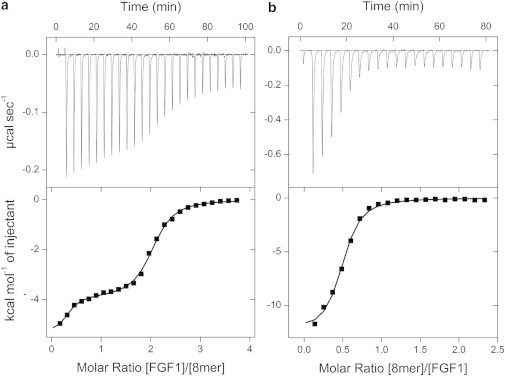
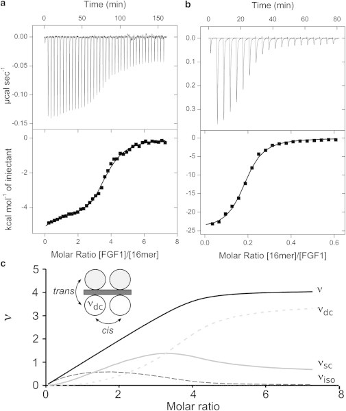
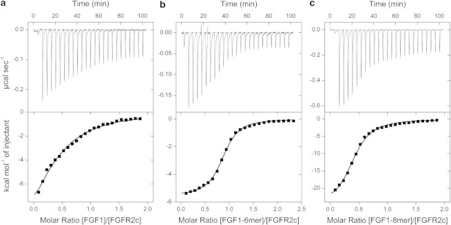
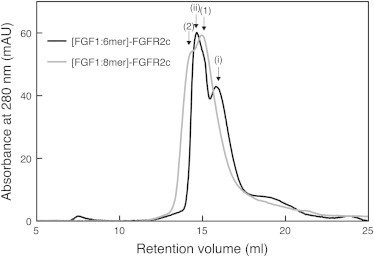
Fibroblast growth factors (FGFs) utilize cell surface heparan sulfate as a coreceptor in the assembly of signaling complexes with FGF-receptors on the plasma membrane. Here we undertake a complete thermodynamic characterization of the assembly of the FGF signaling complex using isothermal titration calorimetry. Heparin fragments of defined length are used as chemical analogs of the sulfated domains of heparan sulfate and examined for their ability to oligomerize FGF1. Binding is modeled using the McGhee-von Hippel formalism for the cooperative binding of ligands to a monodimensional lattice. Oligomerization of FGFs on heparin is shown to be mediated by positive cooperativity (α = 6). Heparin octasaccharide is the shortest length capable of dimerizing FGF1 and on longer heparin chains FGF1 binds with a minimal footprint of 4.2 saccharide units. The thermodynamics and stoichiometry of the ternary complex suggest that in solution FGF1 binds to heparin in a trans-dimeric manner before FGFR recruitment.
Copyright © 2013 Biophysical Society. Published by Elsevier Inc. All rights reserved.
Figures
Similar articles
-
Cooperative dimerization of fibroblast growth factor 1 (FGF1) upon a single heparin saccharide may drive the formation of 2:2:1 FGF1.FGFR2c.heparin ternary complexes.J Biol Chem. 2005 Dec 23;280(51):42274-82. doi: 10.1074/jbc.M505720200. Epub 2005 Oct 11. J Biol Chem. 2005. PMID: 16219767
-
Influence of heparin mimetics on assembly of the FGF.FGFR4 signaling complex.J Biol Chem. 2010 Aug 20;285(34):26628-40. doi: 10.1074/jbc.M109.095109. Epub 2010 Jun 14. J Biol Chem. 2010. PMID: 20547770 Free PMC article.
-
Heparin modulates the mitogenic activity of fibroblast growth factor by inducing dimerization of its receptor. a 3D view by using NMR.Chembiochem. 2013 Sep 23;14(14):1732-44. doi: 10.1002/cbic.201300313. Epub 2013 Aug 12. Chembiochem. 2013. PMID: 23940086
-
Paradigms in the structural biology of the mitogenic ternary complex FGF:FGFR:heparin.Biochimie. 2016 Aug;127:214-26. doi: 10.1016/j.biochi.2016.05.017. Epub 2016 Jun 2. Biochimie. 2016. PMID: 27263122 Review.
-
Role of heparan sulfate in fibroblast growth factor signalling: a structural view.Curr Opin Struct Biol. 2001 Oct;11(5):629-34. doi: 10.1016/s0959-440x(00)00258-x. Curr Opin Struct Biol. 2001. PMID: 11785766 Review.
Cited by
-
Heparan Sulfate Domains Required for Fibroblast Growth Factor 1 and 2 Signaling through Fibroblast Growth Factor Receptor 1c.J Biol Chem. 2017 Feb 10;292(6):2495-2509. doi: 10.1074/jbc.M116.761585. Epub 2016 Dec 28. J Biol Chem. 2017. PMID: 28031461 Free PMC article.
-
Proteoglycans: a common portal for SARS-CoV-2 and extracellular vesicle uptake.Am J Physiol Cell Physiol. 2023 Jan 1;324(1):C76-C84. doi: 10.1152/ajpcell.00453.2022. Epub 2022 Dec 2. Am J Physiol Cell Physiol. 2023. PMID: 36458979 Free PMC article. Review.
-
Roles of glycosaminoglycans as regulators of ligand/receptor complexes.Open Biol. 2018 Oct 3;8(10):180026. doi: 10.1098/rsob.180026. Open Biol. 2018. PMID: 30282658 Free PMC article. Review.
-
Gas-Phase Analysis of the Complex of Fibroblast GrowthFactor 1 with Heparan Sulfate: A Traveling Wave Ion Mobility Spectrometry (TWIMS) and Molecular Modeling Study.J Am Soc Mass Spectrom. 2017 Jan;28(1):96-109. doi: 10.1007/s13361-016-1496-8. Epub 2016 Sep 23. J Am Soc Mass Spectrom. 2017. PMID: 27663556 Free PMC article.
-
The crystal structure of fibroblast growth factor 18 (FGF18).Protein Cell. 2014 May;5(5):343-7. doi: 10.1007/s13238-014-0033-4. Protein Cell. 2014. PMID: 24668462 Free PMC article. No abstract available.
References
-
- Ornitz D.M. FGFs, heparan sulfate and FGFRs: complex interactions essential for development. Bioessays. 2000;22:108–112. - PubMed
-
- Presta M., Dell’Era P., Rusnati M. Fibroblast growth factor/fibroblast growth factor receptor system in angiogenesis. Cytokine Growth Factor Rev. 2005;16:159–178. - PubMed
-
- Powers C.J., McLeskey S.W., Wellstein A. Fibroblast growth factors, their receptors and signaling. Endocr. Relat. Cancer. 2000;7:165–197. - PubMed
-
- Johnson D.E., Williams L.T. Structural and functional diversity in the FGF receptor multigene family. Adv. Cancer Res. 1993;60:1–41. - PubMed
Publication types
MeSH terms
Substances
Grants and funding
LinkOut - more resources
Full Text Sources
Other Literature Sources
Medical
Miscellaneous
