

Multiphasic enhancement patterns of small renal masses (≤4 cm) on preoperative computed tomography: utility for distinguishing subtypes of renal cell carcinoma, angiomyolipoma, and oncocytoma
- PMID: 23601445
- PMCID: PMC4648275
- DOI: 10.1016/j.urology.2012.12.049
Multiphasic enhancement patterns of small renal masses (≤4 cm) on preoperative computed tomography: utility for distinguishing subtypes of renal cell carcinoma, angiomyolipoma, and oncocytoma
Abstract
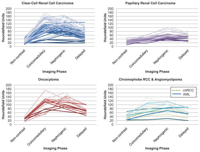
Objective: To analyze the enhancement patterns of small renal masses (SRMs) during 4-phase computed tomography (CT) imaging to predict histology.
Methods: One-hundred consecutive patients with SRMs and 4-phase preoperative CT imaging, who underwent extirpative surgery with a pathologic diagnosis of renal cell carcinoma (RCC), angiomyolipoma (AML), or oncocytoma, were identified from a single institution. An expert radiologist, blinded to histologic results, retrospectively recorded tumor size, RENAL (radius, exophytic/endophytic properties of the tumor, nearness of tumor deepest portion to the collecting system or sinus, anterior/posterior descriptor, and the location relative to polar lines) nephrometry score, tumor attenuation, and the renal cortex on all 4 acquisitions (precontrast, corticomedullary, nephrogenic, and delayed density).
Results: Pathologic diagnoses included 48 clear-cell RCCs (ccRCCs), 22 papillary RCCs, 10 chromophobe RCCs, 13 oncocytomas, and 7 AMLs. There was no significant difference in median tumor size (P = .8), nephrometry score (P = .98), or anatomic location (P >.2) among histologies. Significant differences were noted in peak enhancement (P <.001) and phase-specific enhancement (P <.007) by histology. Papillary RCCs demonstrated a distinct enhancement pattern, with a peak Hounsfield unit (HU) of 56, and greatest enhancement during the NG and delayed phases. The highest peak HU were demonstrated by ccRCC (117 HU) and oncocytoma (125 HU); ccRCC more often peaked in the corticomedullary phase, whereas oncocytoma peaked in the nephrogenic phase.
Conclusion: In a series of patients with SRMs undergoing 4-phase CT, tumor histologies demonstrated distinct enhancement patterns. Thus, preoperative 4-phase CT imaging may provide useful information regarding pathologic diagnosis in patients undergoing extirpative surgery.
Copyright © 2013 Elsevier Inc. All rights reserved.
Figures
Comment in
-
Editorial comment.Urology. 2013 Jun;81(6):1271-2. doi: 10.1016/j.urology.2012.12.052. Epub 2013 Apr 17. Urology. 2013. PMID: 23601440 No abstract available.
-
Kidney cancer: Multiphasic CT to distinguish small renal mass subtype.Nat Rev Urol. 2013 Jun;10(6):310. doi: 10.1038/nrurol.2013.102. Epub 2013 May 7. Nat Rev Urol. 2013. PMID: 23649291 No abstract available.
References
-
- Hollingsworth JM, Miller DC, Daignault S, et al. Rising incidence of small renal masses: a need to reassess treatment effect. J Natl Cancer Inst. 2006;98:1331–1334. - PubMed
-
- Cancer Facts and Figures 2010. Atlanta, GA: The American Cancer Society; 2010. [Accessed September 1, 2011]. http://www.cancer.org/acs/groups/content/@epidemiologysurveilance/docume....
-
- Kane CJ, Mallin K, Ritchey J, et al. Renal cell cancer stage migration: analysis of the National Cancer Data Base. Cancer. 2008;113:78–83. - PubMed
-
- Nguyen MM, Gill IS, Ellison LM. The evolving presentation of renal carcinoma in the United States: trends from the Surveillance, Epidemiology, and End Results program. J Urol. 2006;176:2397–2400. discussion 2400. - PubMed
-
- Kutikov A, Fossett LK, Ramchandani P, et al. Incidence of benign pathologic findings at partial nephrectomy for solitary renal mass presumed to be renal cell carcinoma on preoperative imaging. Urology. 2006;68:737–740. - PubMed
MeSH terms
Substances
Supplementary concepts
Grants and funding
LinkOut - more resources
Full Text Sources
Other Literature Sources
Medical
