

Notch4 is required for tumor onset and perfusion
- PMID: 23601498
- PMCID: PMC3644271
- DOI: 10.1186/2045-824X-5-7
Notch4 is required for tumor onset and perfusion
Abstract
Background: Notch4 is a member of the Notch family of receptors that is primarily expressed in the vascular endothelial cells. Genetic deletion of Notch4 does not result in an overt phenotype in mice, thus the function of Notch4 remains poorly understood.
Methods: We examined the requirement for Notch4 in the development of breast cancer vasculature. Orthotopic transplantation of mouse mammary tumor cells wild type for Notch4 into Notch4 deficient hosts enabled us to delineate the contribution of host Notch4 independent of its function in the tumor cell compartment.
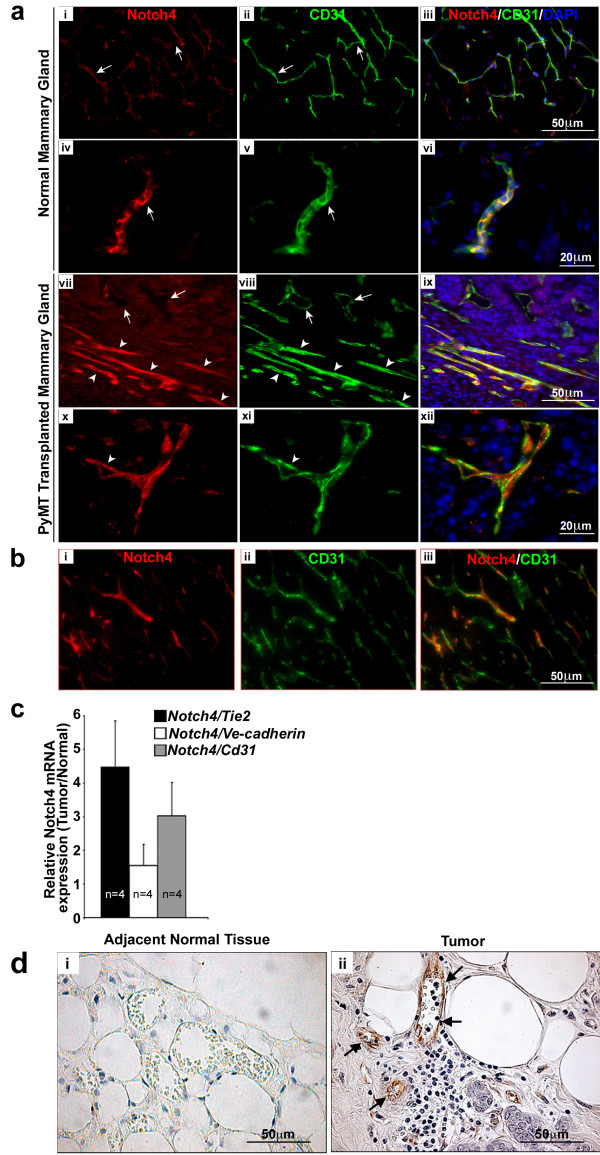
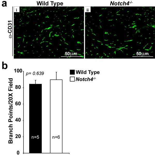
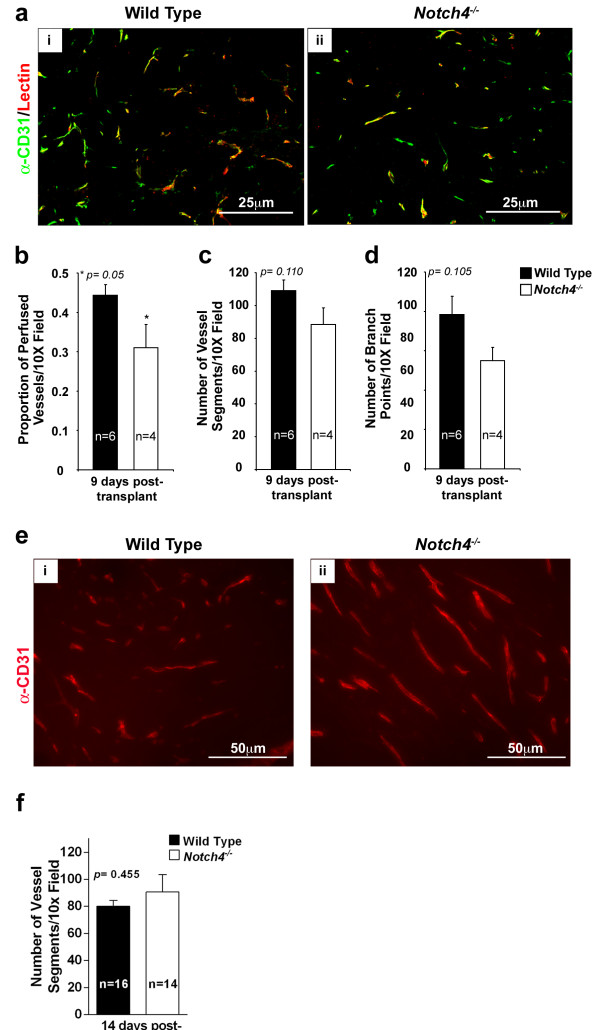
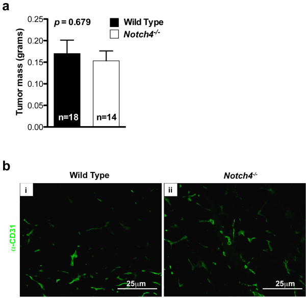
Results: Here, we show that Notch4 expression is required for tumor onset and early tumor perfusion in a mouse model of breast cancer. We found that Notch4 expression is upregulated in mouse and human mammary tumor vasculature. Moreover, host Notch4 deficiency delayed the onset of MMTV-PyMT tumors, wild type for Notch4, after transplantation. Vessel perfusion was decreased in tumors established in Notch4-deficient hosts. Unlike in inhibition of Notch1 or Dll4, vessel density and branching in tumors developed in Notch4-deficient mice were unchanged. However, final tumor size was similar between tumors grown in wild type and Notch4 null hosts.
Conclusion: Our results suggest a novel role for Notch4 in the establishment of tumor colonies and vessel perfusion of transplanted mammary tumors.
Figures

Similar articles
-
The vascular delta-like ligand-4 (DLL4)-Notch4 signaling correlates with angiogenesis in primary glioblastoma: an immunohistochemical study.Tumour Biol. 2016 Mar;37(3):3797-805. doi: 10.1007/s13277-015-4202-8. Epub 2015 Oct 15. Tumour Biol. 2016. PMID: 26472724
-
Inhibition of Notch4 Using Novel Neutralizing Antibodies Reduces Tumor Growth in Murine Cancer Models by Targeting the Tumor Endothelium.Cancer Res Commun. 2024 Jul 1;4(7):1881-1893. doi: 10.1158/2767-9764.CRC-24-0081. Cancer Res Commun. 2024. PMID: 38984877 Free PMC article.
-
Notch4 reveals a novel mechanism regulating Notch signal transduction.Biochim Biophys Acta. 2014 Jul;1843(7):1272-84. doi: 10.1016/j.bbamcr.2014.03.015. Epub 2014 Mar 22. Biochim Biophys Acta. 2014. PMID: 24667410
-
Repression of the putative tumor suppressor gene Bard1 or expression of Notch4(int-3) oncogene subvert the morphogenetic properties of mammary epithelial cells.Adv Exp Med Biol. 2000;480:175-84. doi: 10.1007/0-306-46832-8_22. Adv Exp Med Biol. 2000. PMID: 10959425 Review.
-
Notch in mammary gland development and breast cancer.Semin Cancer Biol. 2004 Oct;14(5):341-7. doi: 10.1016/j.semcancer.2004.04.013. Semin Cancer Biol. 2004. PMID: 15288259 Review.
Cited by
-
Notch1 and Notch4 core binding domain peptibodies exhibit distinct ligand-binding and anti-angiogenic properties.Angiogenesis. 2023 May;26(2):249-263. doi: 10.1007/s10456-022-09861-6. Epub 2022 Nov 15. Angiogenesis. 2023. PMID: 36376768 Free PMC article.
-
Unique functions for Notch4 in murine embryonic lymphangiogenesis.Angiogenesis. 2022 May;25(2):205-224. doi: 10.1007/s10456-021-09822-5. Epub 2021 Oct 19. Angiogenesis. 2022. PMID: 34665379 Free PMC article.
-
AKT and 14-3-3 regulate Notch4 nuclear localization.Sci Rep. 2015 Mar 5;5:8782. doi: 10.1038/srep08782. Sci Rep. 2015. PMID: 25740432 Free PMC article.
-
Targeting Notch4 in Cancer: Molecular Mechanisms and Therapeutic Perspectives.Cancer Manag Res. 2021 Sep 8;13:7033-7045. doi: 10.2147/CMAR.S315511. eCollection 2021. Cancer Manag Res. 2021. PMID: 34526819 Free PMC article. Review.
-
Delta-like ligand 4 (DLL4) in the plasma and neoplastic tissues from breast cancer patients: correlation with metastasis.Med Oncol. 2014 May;31(5):945. doi: 10.1007/s12032-014-0945-0. Epub 2014 Apr 3. Med Oncol. 2014. PMID: 24696220
References
-
- Uyttendaele H, Marazzi G, Wu G, Yan Q, Sassoon D, Kitajewski J. Notch4/int-3, a mammary proto-oncogene, is an endothelial cell-specific mammalian Notch gene. Development. 1996;122(7):2251–2259. - PubMed
LinkOut - more resources
Full Text Sources
Other Literature Sources
