

Why does the healthy cornea resist Pseudomonas aeruginosa infection?
- PMID: 23601656
- PMCID: PMC3718454
- DOI: 10.1016/j.ajo.2013.03.001
Why does the healthy cornea resist Pseudomonas aeruginosa infection?
Abstract
Purpose: To provide our perspective on why the cornea is resistant to infection based on our research results with Pseudomonas (P) aeruginosa. We focus on our current understanding of the interplay between bacteria, tear fluid, and the corneal epithelium that determines health as the usual outcome, and propose a theoretical model for how contact lens wear might change those interactions to enable susceptibility to P aeruginosa infection.
Methods: Use of "null-infection" in vivo models, cultured human corneal epithelial cells, contact lens-wearing animal models, and bacterial genetics help to elucidate mechanisms by which P aeruginosa survives at the ocular surface, adheres, and traverses multilayered corneal epithelia. These models also help elucidate the molecular mechanisms of corneal epithelial innate defense.
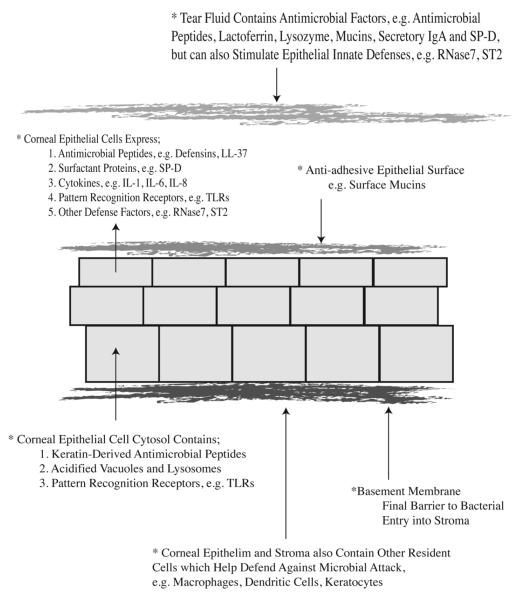
Results: Tear fluid and the corneal epithelium combine to make a formidable defense against P aeruginosa infection of the cornea. Part of that defense involves the expression of antimicrobials such as β-defensins, the cathelicidin LL-37, cytokeratin-derived antimicrobial peptides, and RNase7. Immunomodulators such as SP-D and ST2 also contribute. Innate defenses of the cornea depend in part on MyD88, a key adaptor protein of TLR and IL-1R signaling, but the basal lamina represents the final barrier to bacterial penetration. Overcoming these defenses involves P aeruginosa adaptation, expression of the type III secretion system, proteases, and P aeruginosa biofilm formation on contact lenses.
Conclusion: After more than 2 decades of research focused on understanding how contact lens wear predisposes to P aeruginosa infection, our working hypothesis places blame for microbial keratitis on bacterial adaptation to ocular surface defenses, combined with changes to the biochemistry of the corneal surface caused by trapping bacteria and tear fluid against the cornea under the lens.
Copyright © 2013 Elsevier Inc. All rights reserved.
Figures
References
-
- Lichtinger A, Yeung SN, Kim P, et al. Shifting trends in bacterial keratitis in toronto: an 11-year review. Ophthalmology. 2012;119(9):1785–1790. - PubMed
-
- Green M, Apel A, Stapleton F. Risk factors and causative organisms in microbial keratitis. Cornea. 2008;27(1):22–27. - PubMed
-
- Hazlett LD, Hendricks RL. Reviews for immune privilege in the year 2010: immune privilege and infection. Ocul Immunol Inflamm. 2010;18(4):237–243. - PubMed
-
- Hazlett LD. Bacterial infections of the cornea (Pseudomonas aeruginosa) Chem Immunol Allergy. 2007;92:185–194. - PubMed
Publication types
MeSH terms
Substances
Grants and funding
LinkOut - more resources
Full Text Sources
Other Literature Sources
Medical
Research Materials
