Mimicking white matter tract topography using core-shell electrospun nanofibers to examine migration of malignant brain tumors
- PMID: 23601662
- PMCID: PMC4080638
- DOI: 10.1016/j.biomaterials.2013.03.069
Mimicking white matter tract topography using core-shell electrospun nanofibers to examine migration of malignant brain tumors
Abstract
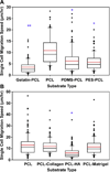
Glioblastoma multiforme (GBM), one of the deadliest forms of human cancer, is characterized by its high infiltration capacity, partially regulated by the neural extracellular matrix (ECM). A major limitation in developing effective treatments is the lack of in vitro models that mimic features of GBM migration highways. Ideally, these models would permit tunable control of mechanics and chemistry to allow the unique role of each of these components to be examined. To address this need, we developed aligned nanofiber biomaterials via core-shell electrospinning that permit systematic study of mechanical and chemical influences on cell adhesion and migration. These models mimic the topography of white matter tracts, a major GBM migration 'highway'. To independently investigate the influence of chemistry and mechanics on GBM behaviors, nanofiber mechanics were modulated by using different polymers (i.e., gelatin, poly(ethersulfone), poly(dimethylsiloxane)) in the 'core' while employing a common poly(ε-caprolactone) (PCL) 'shell' to conserve surface chemistry. These materials revealed GBM sensitivity to nanofiber mechanics, with single cell morphology (Feret diameter), migration speed, focal adhesion kinase (FAK) and myosin light chain 2 (MLC2) expression all showing a strong dependence on nanofiber modulus. Similarly, modulating nanofiber chemistry using extracellular matrix molecules (i.e., hyaluronic acid (HA), collagen, and Matrigel) in the 'shell' material with a common PCL 'core' to conserve mechanical properties revealed GBM sensitivity to HA; specifically, a negative effect on migration. This system, which mimics the topographical features of white matter tracts, should allow further examination of the complex interplay of mechanics, chemistry, and topography in regulating brain tumor behaviors.
Copyright © 2013 Elsevier Ltd. All rights reserved.
Figures


Similar articles
-
Glioma-astrocyte interactions on white matter tract-mimetic aligned electrospun nanofibers.Biotechnol Prog. 2015 Sep-Oct;31(5):1406-15. doi: 10.1002/btpr.2123. Epub 2015 Jun 26. Biotechnol Prog. 2015. PMID: 26081199
-
Polycaprolactone/Gelatin/Hyaluronic Acid Electrospun Scaffolds to Mimic Glioblastoma Extracellular Matrix.Materials (Basel). 2020 Jun 11;13(11):2661. doi: 10.3390/ma13112661. Materials (Basel). 2020. PMID: 32545241 Free PMC article.
-
Bioengineered 3D brain tumor model to elucidate the effects of matrix stiffness on glioblastoma cell behavior using PEG-based hydrogels.Mol Pharm. 2014 Jul 7;11(7):2115-25. doi: 10.1021/mp5000828. Epub 2014 Apr 29. Mol Pharm. 2014. PMID: 24712441
-
Recent advances in brain tumor therapy: application of electrospun nanofibers.Drug Discov Today. 2018 Apr;23(4):912-919. doi: 10.1016/j.drudis.2018.02.007. Epub 2018 Feb 27. Drug Discov Today. 2018. PMID: 29499377 Review.
-
Glioblastoma mechanobiology at multiple length scales.Biomater Adv. 2024 Jun;160:213860. doi: 10.1016/j.bioadv.2024.213860. Epub 2024 Apr 15. Biomater Adv. 2024. PMID: 38640876 Review.
Cited by
-
Effect of Electrospun Fiber Mat Thickness and Support Method on Cell Morphology.Nanomaterials (Basel). 2019 Apr 20;9(4):644. doi: 10.3390/nano9040644. Nanomaterials (Basel). 2019. PMID: 31010029 Free PMC article.
-
Functionalized Antimicrobial Nanofibers: Design Criteria and Recent Advances.J Funct Biomater. 2021 Oct 28;12(4):59. doi: 10.3390/jfb12040059. J Funct Biomater. 2021. PMID: 34842715 Free PMC article. Review.
-
Biomimetic Approach of Brain Vasculature Rapidly Characterizes Inter- and Intra-Patient Migratory Diversity of Glioblastoma.Small Methods. 2024 Dec;8(12):e2400210. doi: 10.1002/smtd.202400210. Epub 2024 May 15. Small Methods. 2024. PMID: 38747088 Free PMC article.
-
The scrambled story between hyaluronan and glioblastoma.J Biol Chem. 2021 Jan-Jun;296:100549. doi: 10.1016/j.jbc.2021.100549. Epub 2021 Mar 17. J Biol Chem. 2021. PMID: 33744285 Free PMC article. Review.
-
Dissecting and rebuilding the glioblastoma microenvironment with engineered materials.Nat Rev Mater. 2019 Oct;4(10):651-668. doi: 10.1038/s41578-019-0135-y. Epub 2019 Aug 16. Nat Rev Mater. 2019. PMID: 32647587 Free PMC article.
References
-
- Wen PY, Kesari S. Malignant gliomas in adults. N Engl J Med. 2008;359:492–507. - PubMed
-
- Bao SD, Wu QL, McLendon RE, Hao YL, Shi Q, Hjelmeland AB, et al. Glioma stem cells promote radioresistance by preferential activation of the DNA damage response. Nature. 2006;444:756–760. - PubMed
-
- Sarkar A, Chiocca EA. Glioblastoma and malignant astrocytoma. In: Laws Ka., editor. Brain tumors: an encyclopedic approach. 3rd ed. Edinburgh, New York, USA: Churchill Livingstone; 2011.
-
- Louis DN. Molecular pathology of malignant gliomas. Annu Rev Pathol. 2006;1:97–117. - PubMed
Publication types
MeSH terms
Substances
Grants and funding
LinkOut - more resources
Full Text Sources
Other Literature Sources
Medical
Miscellaneous

