. 2013 Sep;34(9):2146-57.
doi: 10.1016/j.neurobiolaging.2013.03.015. Epub 2013 Apr 17.

Tau phosphorylation affects its axonal transport and degradation

Affiliations

Tau phosphorylation affects its axonal transport and degradation

Teresa Rodríguez-Martín et al. Neurobiol Aging. 2013 Sep.

Abstract

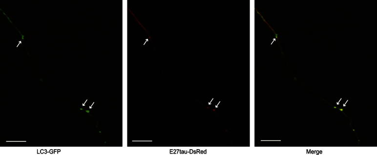
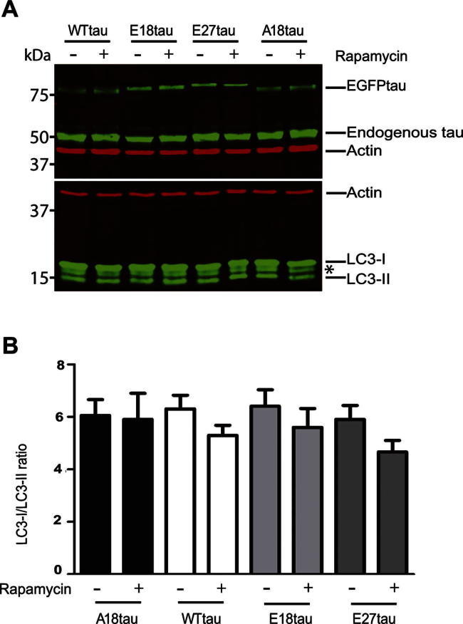
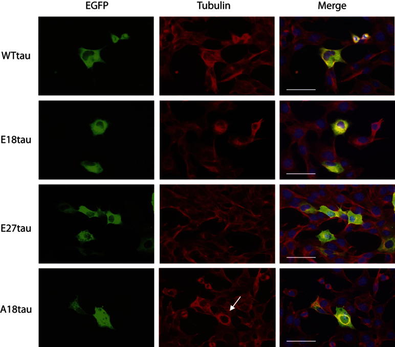
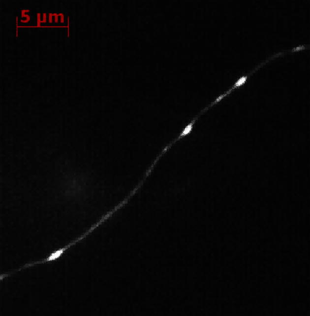
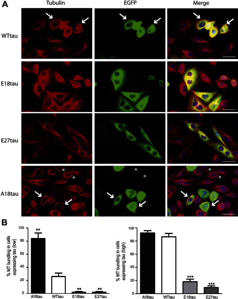
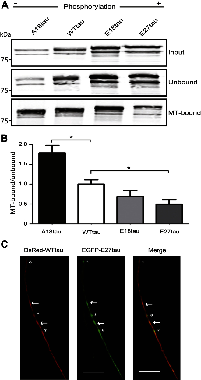
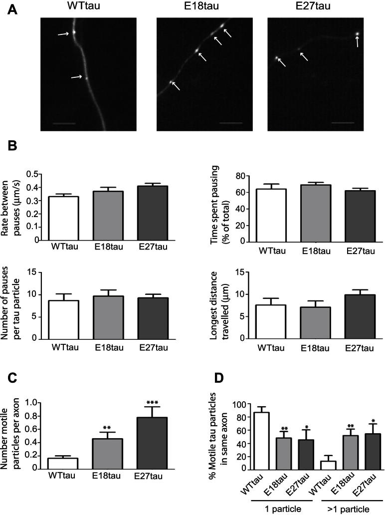
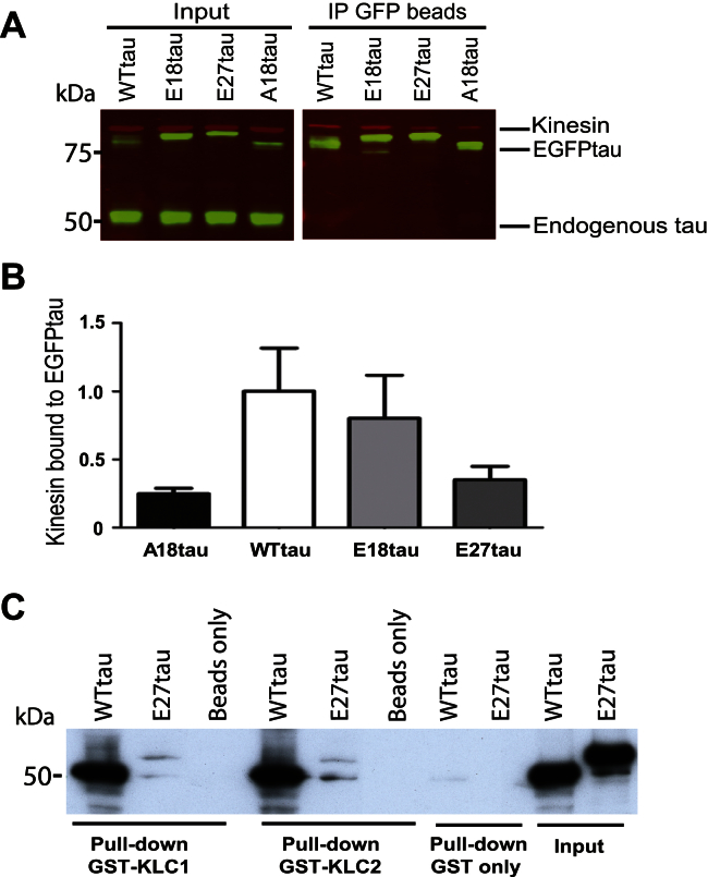
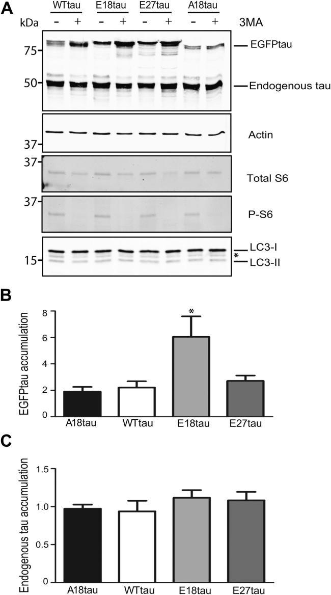
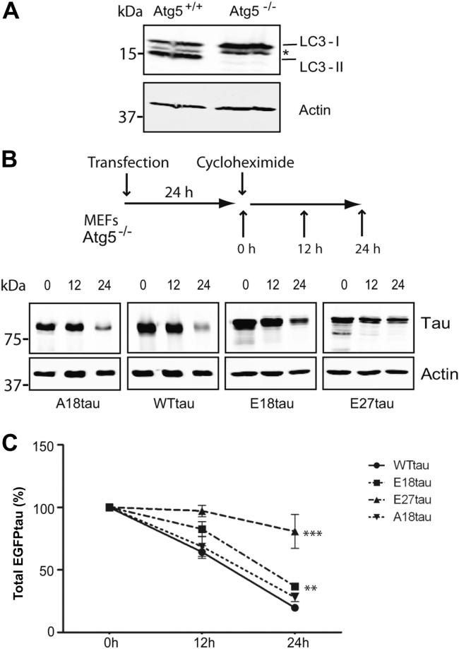
Phosphorylated forms of microtubule-associated protein tau accumulate in neurofibrillary tangles in Alzheimer's disease. To investigate the effects of specific phosphorylated tau residues on its function, wild type or phosphomutant tau was expressed in cells. Elevated tau phosphorylation decreased its microtubule binding and bundling, and increased the number of motile tau particles, without affecting axonal transport kinetics. In contrast, reducing tau phosphorylation enhanced the amount of tau bound to microtubules and inhibited axonal transport of tau. To determine whether differential tau clearance is responsible for the increase in phosphomimic tau, we inhibited autophagy in neurons which resulted in a 3-fold accumulation of phosphomimic tau compared with wild type tau, and endogenous tau was unaffected. In autophagy-deficient mouse embryonic fibroblasts, but not in neurons, proteasomal degradation of phosphomutant tau was also reduced compared with wild type tau. Therefore, autophagic and proteasomal pathways are involved in tau degradation, with autophagy appearing to be the primary route for clearing phosphorylated tau in neurons. Defective autophagy might contribute to the accumulaton of tau in neurodegenerative diseases.


Figures

Supplementary Fig. 1
Colocalization of E27tau with LC3. Still image of neurons transfected with LC3-GFP and E27tau-DsRed. After 48 hours, time-lapse movies showed colocalization of E27tau with LC3 (arrows). Scale bar, 5 μm. Abbreviations: GFP, green fluorescent protein; LC3, light-chain 3.
Supplementary Fig. 2
Lack of effect of rapamycin on tau degradation in neurons. Neurons were transfected with each tau construct and after 48-hour expression the cells were treated for 24 hours with 200 nM rapamycin to activate autophagy. (A) Total cell lysates were probed on Western blots using antibodies against tau (green), β-actin (red), and LC3 (green). * Nonspecific band detected by the LC3 antibody. (B) LC3-I/LC3-II was calculated for each condition (mean ± standard error of the mean; n = 3). Rapamycin did not increase LC3-II or reduce the LC3-I/LC3-II ratio as would be expected after activation of autophagy. Abbreviation: LC3, light-chain 3.
Supplementary Fig. 3
Wild type and phosphomutant tau species do not aggregate in autophagy-deficient MEFs. WTtau, E18tau, E27tau, and A18tau were expressed in transfected autophagy-deficient (Atg5-/-) MEFs. Tubulin was stained using DM1A antibody and tau was detected by EGFP fluorescence. No tau aggregates were detected after 48-hour expression. A18tau exhibited a greater ability to bundle microtubules (white arrow). Scale bar, 50 μm. Abbreviations: EGFP, enhanced green fluorescent protein; MEF, mouse embryonic fibroblast cell.
Supplementary Fig. 4
Supplementary Fig. 5
Supplementary Fig. 6
Fig. 1
Fig. 1
Tau phosphomutants. The diagram shows the positions in the longest human tau isoform (2N4R, 441 residues) of the serine (S) or threonine (T) residues mutated to glutamate (to mimic phosphorylation) in E18tau and E27tau, or to alanine (to preclude phosphorylation) in A18tau. R1, R2, R3, and R4 are the repeat regions of the microtubule (MT)-binding domain.
Fig. 2
Fig. 2
Microtubule bundling of tau constructs. (A) CHO cells were transfected with WTtau, E18tau, E27tau, or A18tau and tau expression was visualized by EGFP fluorescence. Microtubules were labeled using the monoclonal α-tubulin antibody, DM1A. WTtau and A18tau exhibited a greater ability to bundle microtubules (arrows). Asterisks indicate cells with microtubule bundles in the presence of low amounts of A18tau. Scale bar, 50μm. (B) Quantification of microtubule-bundling in cells expressing high or low amounts of tau. Values shown are mean ± standard error of the mean. ** p < 0.01; *** p < 0.001, Student t test, relative to WTtau. Abbreviations: CHO, Chinese hamster ovary; EGFP, enhanced green fluorescent protein; MT, microtubule.
Fig. 3
Fig. 3
Microtubule binding of wild type and phosphomutant tau. (A) CHO cells transfected with A18tau, WTtau, E18tau, and E27tau were separated into microtubule-bound and unbound fractions and analyzed using Western blot using an antibody to tau (DAKO). (B) Quantification of microtubule-bound/unbound expressed relative to WTtau (mean ± standard error of the mean; n = 3); * p < 0.05, Student t test, relative to WTtau. (C) Coexpression of DsRed-tagged WTtau and EGFP-tagged E27tau in rat cortical neurons. Arrows indicate colocalization of WTtau and E27tau and asterisks indicate individual E27tau particles. Scale bar, 10 μm. Abbreviations: CHO, Chinese hamster ovary; EGFP, enhanced green fluorescent protein; MT, microtubule.
Fig. 4
Fig. 4
Fast axonal transport of tau. (A) WTtau, E18tau, and E27tau exhibit a punctate morphology in axons in transfected neurons. Scale bar, 5 μm. (B) Kinetic parameters of motile tau particles. Rate between pauses, time spent pausing, number of pauses, and longest distance traveled between pauses were calculated for each tau construct (WTtau, n = 154; E18tau, n = 89; E27tau, n = 81). (C) The number of motile particles per axon is shown for each tau construct. (D) The probability of finding 1 or more than 1 motile tau particle moving in an individual axon was calculated from a randomized selection of movies (WTtau, n =12; E18tau, n = 8; E27tau, n = 7). Values shown are mean ± standard error of the mean. * p < 0.05, ** p <0.01, *** p < 0.001, Student t test, relative to WTtau.
Fig. 5
Fig. 5
Tau pseudophosphorylation does not influence its binding to kinesin. (A) WTtau, E18tau, E27tau, or A18tau were expressed in primary rat cortical neurons and immunoprecipitated (IP) using an antibody to GFP. Total input and IP fractions were analyzed on Western blots probed with antibodies to kinesin heavy chain (red) and tau (green). (B) Kinesin/EGFPtau for each tau construct is expressed relative to WTtau. Values shown are mean ± standard error of the mean; n = 3; Student t test, relative to WTtau, showed no statistically significant differences. (C) Western blot of GST pull-down of WTtau and E27tau with GST-KLC1 and GST-KLC2, probed with tau antibody (TP70). Abbreviations: EGFP, enhanced green fluorescent protein; GFP, green fluorescent protein; GST, glutathione-S-transferase; KLC, kinesin light chain.
Fig. 6
Fig. 6
Inhibiting autophagy in cortical neurons results in differential degradation of tau phospho-mutants. Neurons were transfected with each tau construct and treated with 10 mM 3MA. (A) Cell lysates were analyzed on Western blots probed with antibodies to tau, β-actin, total and phosphorylated (P) ribosomal S6 protein, and LC3. * Nonspecific band detected by the LC3 antibody. (B) EGFPtau accumulation expressed as a ratio of 3MA-treated and untreated neurons, normalized to β-actin. Values shown are mean ± SEM; n = 5–6; 1-way analysis of variance. (C) Endogenous tau accumulation expressed as a ratio of 3MA-treated and untreated neurons, normalized to β-actin. Values shown are mean ± SEM; n = 6. Abbreviations: EGFP, enhanced green fluorescent protein; LC3, light chain 3; SEM, standard error of the mean.
Fig. 7
Fig. 7
Autophagy-deficient MEFs exhibit differential degradation of tau phospho-mutants. (A) Western blot of Atg5+/+ and Atg5-/- MEFs probed with antibodies to LC3 and β-actin. * Nonspecific band detected by the LC3 antibody. (B) Atg5-/- MEFs were transfected with each tau construct. After 24 hours, cells were treated with 100 μM cycloheximide and harvested at 0, 12, and 24 hours. Cell lysates were analyzed on Western blots probed with antibodies to tau (DAKO) and β-actin. (C) The amount of tau remaining after cycloheximide treatment for 12 and 24 hours was calculated as the percentage of tau present at 0 hours. Values shown are mean ± standard error of the mean; n = 6; ** p < 0.01, *** p < 0.001, Student t test, relative to WTtau at 24 hours. Abbreviations: EGFP, enhanced green fluorescent protein; LC3, light chain 3; MEF, mouse embryonic fibroblasts.
Fig. 8
Fig. 8
Lack of tau degradation by the proteasome in neurons. (A) Primary rat cortical neurons were transfected with each tau construct. After 48 hours, neurons were treated with 100 μM cycloheximide and harvested at 0, 12, and 24 hours. Neuronal lysates were analyzed on Western blots probed with antibodies to tau (DAKO) and β-actin. (B) The amount of EGFPtau remaining after cycloheximide treatment for 12 and 24 hours was calculated as the percentage of tau present at 0 hours. Values shown are mean ± standard error of the mean; n = 6. Abbreviations: DIV, days in vitro; EGFP, enhanced green fluorescent protein; Endog, endogenous.

Similar articles

Cited by

References

    1. Ackerley S., Thornhill P., Grierson A.J., Brownlees J., Anderton B.H., Leigh P.N., Shaw C.E., Miller C.C. Neurofilament heavy chain side arm phosphorylation regulates axonal transport of neurofilaments. J. Cell Biol. 2003;161:489–495. - PMC - PubMed
    1. Aplin A., Jasionowski T., Tuttle D.L., Lenk S.E., Dunn W.A., Jr. Cytoskeletal elements are required for the formation and maturation of autophagic vacuoles. J. Cell Physiol. 1992;152:458–466. - PubMed
    1. Avila J., Leon-Espinosa G., Garcia E., Garcia-Escudero V., Hernandez F., Defelipe J. Tau phosphorylation by GSK3 in different conditions. Int. J. Alzheimers Dis. 2012;2012:578373. - PMC - PubMed
    1. Ballatore C., Lee V.M., Trojanowski J.Q. Tau-mediated neurodegeneration in Alzheimer's disease and related disorders. Nat. Rev. Neurosci. 2007;8:663–672. - PubMed
    1. Bauereis B., Haskins W.E., Lebaron R.G., Renthal R. Proteomic insights into the protective mechanisms of an in vitro oxidative stress model of early stage Parkinson's disease. Neurosci. Lett. 2011;488:11–16. - PMC - PubMed

Publication types

MeSH terms

Substances