Case Reports
. 2013 Jul;120(7):1324-36.
doi: 10.1016/j.ophtha.2012.12.044. Epub 2013 Apr 16.

Ocular manifestations of xeroderma pigmentosum: long-term follow-up highlights the role of DNA repair in protection from sun damage

Affiliations
Case Reports

Ocular manifestations of xeroderma pigmentosum: long-term follow-up highlights the role of DNA repair in protection from sun damage

Brian P Brooks et al. Ophthalmology. 2013 Jul.

Abstract

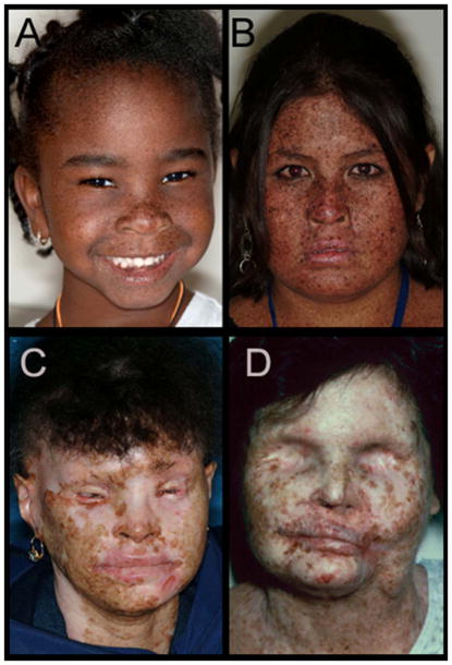
Objective: Xeroderma pigmentosum (XP) is a rare autosomal recessive disease caused by mutations in DNA repair genes. Clinical manifestations of XP include mild to extreme sensitivity to ultraviolet radiation resulting in inflammation and neoplasia in sun-exposed areas of the skin, mucous membranes, and ocular surfaces. This report describes the ocular manifestations of XP in patients systematically evaluated in the Clinical Center at the National Institutes of Health.

Design: Retrospective observational case series.

Participants: Eighty-seven participants, aged 1.3 to 63.4 years, referred to the National Eye Institute (NEI) for examination from 1964 to 2011. Eighty-three patients had XP, 3 patients had XP/Cockayne syndrome complex, and 1 patient had XP/trichothiodystrophy complex.

Methods: Complete age- and developmental stage-appropriate ophthalmic examination.

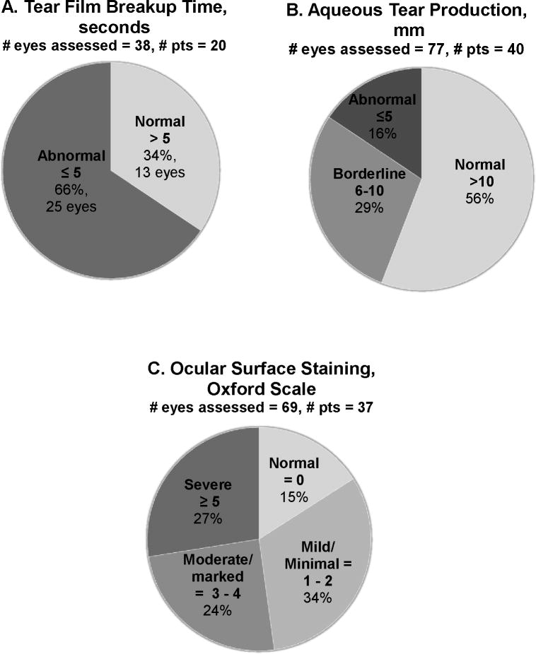
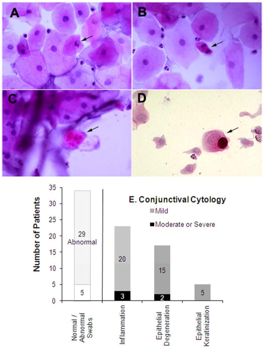
Main outcome measures: Visual acuity; eyelid, ocular surface, and lens pathology; tear film and tear production measures; and cytologic analysis of conjunctival surface swabs.

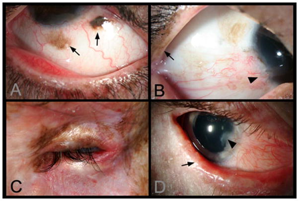
Results: Of the 87 patients, 91% had at least 1 ocular abnormality. The most common abnormalities were conjunctivitis (51%), corneal neovascularization (44%), dry eye (38%), corneal scarring (26%), ectropion (25%), blepharitis (23%), conjunctival melanosis (20%), and cataracts (14%). Thirteen percent of patients had some degree of visual axis impingement, and 5% of patients had no light perception in 1 or both eyes. Ocular surface cancer or a history of ocular surface cancer was present in 10% of patients. Patients with an acute sunburning skin phenotype were less likely to develop conjunctival melanosis and ectropion but more likely to develop neoplastic ocular surface lesions than nonburning patients. Some patients also showed signs of limbal stem cell deficiency.

Conclusions: Our longitudinal study reports the ocular status of the largest group of patients with XP systematically examined at 1 facility over an extended period of time. Structural eyelid abnormalities, neoplasms of the ocular surface and eyelids, tear film and tear production abnormalities, ocular surface disease and inflammation, and corneal abnormalities were present in this population. Burning and nonburning patients with XP exhibit different rates of important ophthalmologic findings, including neoplasia. In addition, ophthalmic characteristics can help refine diagnoses in the case of XP complex phenotypes. DNA repair plays a major role in protection of the eye from sunlight-induced damage.


Conflict of interest statement

Conflict of Interest(s): The authors have no conflicts of interest to declare.

Figures

Figure 1
Figure 1. Facial lentigines and ocular abnormalities in Xeroderma pigmentosum (XP) patients of different ethnicities
A) Case 1 is 5-year old African American XP-C patient (XP444BE) with lentigines on her skin. B) Case 2 is 13-year old First Nations XP-C patient (XP83BE) with many lentigines, ocular surface injection, and sunburn. C) Case 3 is 39-year old Hispanic XP-C patient (XP131BE). She is originally from the Dominican Republic. The patient has a history of skin cancer, but no history of eyelid or ocular surface cancer. Many prominent lentigines are seen on her face and neck. The patient was fairly darkly pigmented when younger, however now pigmentation has been lost, resulting in hypopigmented areas. D) Case 4 is 46-year old Caucasian XP-C patient (XP1BE). She underwent bilateral orbital exenteration at ages 32 and 36 due to recurring invasive squamous cell carcinoma of the ocular surfaces and basal cell carcinoma of the lids, including one squamous cell carcinoma that occurred on the palpebral conjunctiva of the upper eyelid—a non-sun-exposed area. This patient, originally light-skinned, has many darkly pigmented lentiginous areas on her face. She died at age 49 from uterine cancer.,
Figure 2
Figure 2. Lid and ocular surface manifestations in Xeroderma pigmentosum (XP) patients
A) Conjunctival melanosis (arrows) in Case 5, 8-year old Asian Indian XP-C patient (XP417BE). Note the feeder vessels to lesions (arrows). B) Early pterygium (arrowhead) and lid pigmentation (arrow) in Case 2. C) Severe ectropion, entropion, and ocular inflammation in Case 3. D) Lid margin keratinisation (arrow), loss of lashes in Case 6, a 14 year old patient (XP243BE). The patient has a history of skin cancer but no history of ocular surface cancer. Lentigines are present on eyelids and patient has bilateral pterygium, and ectropion. Patient has decreased best corrected visual acuity, possibly due to amblyopia. Localized corneal clouding at leading edge of pterygium was suspicious for early malignancy, biopsy recommended.
Figure 5
Figure 5. High frequency of abnormalities of the tear film, tear production, and ocular surface damage in Xeroderma pigmentosum (XP)-affected patient eyes
A) Tear film breakup times (TBUTs) ≤5 seconds are considered abnormal. No patients had TBUT measured over multiple visits. B) Baseline aqueous tear production measured by Schirmer test II. Schirmer test II values > 10 mm/5 minutes (min) indicate normal baseline tear production, values from 6 mm/5 min to 10 mm/5 min is considered borderline aqueous tear deficiency, and ≤ 5 mm/5 min confirms an aqueous-deficient dry eye categorization. C) Quantification of ocular surface damage in XP-affected patients via epithelial surface staining with fluorescein. The state of the ocular surface was assessed in each patient able to undergo the procedure and graded according to the Oxford Scale. Because not all patients were able to cooperate with this testing, data are available on only a subset of patients.
Figure 6
Figure 6. Conjunctival and corneal surface swabs show severity of ocular surface disease in Xeroderma pigmentosum (XP)-affected patients
Panels A–C show conjunctival swab cytology from Case 3, a 39 year old XP-C patient. Exam discloses many edematous epithelial cells and few acute inflammatory cells. A few epithelial cells are pyknotic. A) A degenerative epithelial cell with a pyknotic nucleus (arrow). B) A mitotic epithelial cell (arrow), C) A degenerative epithelial cell with many cytoplasmic inclusions (arrow). D) Cytology of cells obtained by corneal swab from Case 7 at age 19 (also shown in Figure 3). Degenerative and folded epithelial cells are evident, as well as a mucin-containing goblet cell (arrow), indicative of limbal stem cell deficiency. E) Summary of cytology findings.

References

    1. Kraemer KH, Patronas NJ, Schiffmann R, et al. Xeroderma pigmentosum, trichothiodystrophy and Cockayne syndrome: a complex genotype-phenotype relationship. Neuroscience. 2007;145:1388–96. - PMC - PubMed
    1. Kraemer KH, Lee MM, Scotto J. Xeroderma pigmentosum. Cutaneous, ocular, and neurologic abnormalities in 830 published cases. Arch Dermatol. 1987;123:241–50. - PubMed
    1. Kleijer WJ, Laugel V, Berneburg M, et al. Incidence of DNA repair deficiency disorders in western Europe: xeroderma pigmentosum, Cockayne syndrome and trichothiodystrophy. DNA Repair (Amst) 2008;7:744–50. - PubMed
    1. Neel JV, Kodani M, Brewer R, Anderson RC. The incidence of consanguineous matings in Japan, with remarks on the estimation of comparative gene frequencies and the expected rate of appearance of induced recessive mutations. Am J Hum Genet. 1949;1:156–78. - PMC - PubMed
    1. Kraemer KH, Lee MM, Andrews AD, Lambert WC. The role of sunlight and DNA repair in melanoma and nonmelanoma skin cancer. The xeroderma pigmentosum paradigm. Arch Dermatol. 1994;130:1018–21. - PubMed

Publication types

MeSH terms