

Comparison of automated and manual FISH for evaluation of HER2 gene status on breast carcinoma core biopsies
- PMID: 23601823
- PMCID: PMC3644269
- DOI: 10.1186/1472-6890-13-13
Comparison of automated and manual FISH for evaluation of HER2 gene status on breast carcinoma core biopsies
Abstract
Background: Positive HER2 status identifies breast carcinomas that might respond to trastuzumab treatment. Manual HER2 fluorescent in situ hybridisation (FISH) is the most readily used method to detect HER2 gene amplification which defines positive HER2 status in addition to HER2 protein overexpression. Automation of HER2 FISH may improve HER2 gene testing. The aim of our study was to evaluate an automated HER2 FISH assay for assessing the HER2 genomic status.
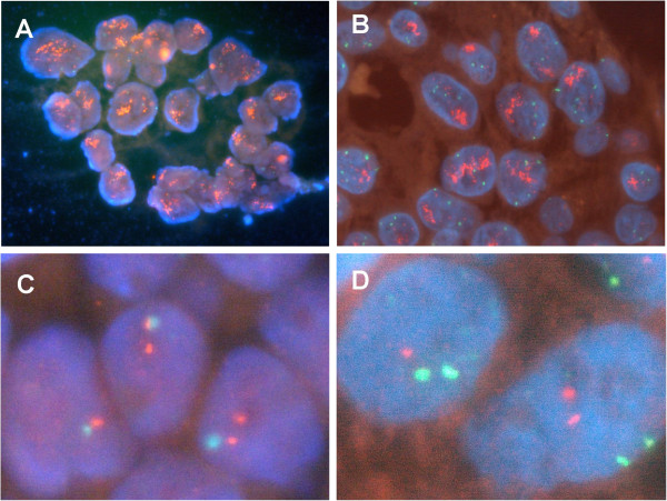
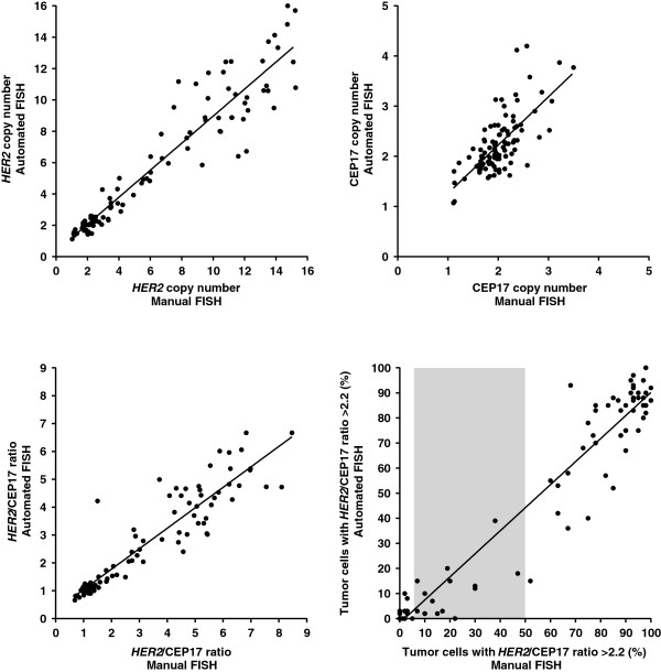
Methods: Core biopsies of 100 invasive breast carcinomas were analysed in parallel using the PathVysion™ HER-2 DNA Probe Kit and the Leica HER2 FISH System for BOND™. To assess inter-method agreement, concordance analysis was performed for various numerical and categorical HER2/CEP17 FISH parameters.
Results: Carcinomas with all HER2 immunohistochemical scores were included (0+: 20; 1+: 20; 2+: 30; 3+: 30). Using either HER2/CEP17 ratio >2.2 or ≥2.0 as criterion for HER2 amplification, high levels of concordance were observed between automated and manual FISH (concordance rate 96%, κ coefficient 0.92). High levels of inter-method agreement were also found for HER2 copy number, CEP17 copy number, HER2/CEP17 ratio, the percentage of carcinoma cells with HER2/CEP17 ratio >2.2, and the presence of HER2 genetic heterogeneity, HER2 clusters and CEP17 polyploidy.
Conclusions: HER2 testing using automated FISH is feasible on breast carcinoma core biopsies. Automated HER2 FISH using the Leica HER2 FISH System for BOND is a practical and efficient alternative to manual HER2 FISH in evaluating the HER2 status of primary invasive breast carcinomas.
Figures
Similar articles
-
Analysis of HER2 status in breast carcinoma by fully automated HER2 fluorescence in situ hybridization (FISH): comparison of two immunohistochemical tests and manual FISH.APMIS. 2014 Sep;122(9):755-60. doi: 10.1111/apm.12215. Epub 2013 Dec 20. APMIS. 2014. PMID: 24372629
-
Evaluation of the HER2 amplification status in oesophageal adenocarcinoma by conventional and automated FISH: a tissue microarray study.J Clin Pathol. 2014 Jan;67(1):26-32. doi: 10.1136/jclinpath-2013-201570. Epub 2013 Sep 16. J Clin Pathol. 2014. PMID: 24043715
-
Automated processing of fluorescence in-situ hybridization slides for HER2 testing in breast and gastro-esophageal carcinomas.Exp Mol Pathol. 2014 Aug;97(1):116-9. doi: 10.1016/j.yexmp.2014.06.003. Epub 2014 Jun 11. Exp Mol Pathol. 2014. PMID: 24927872
-
Evaluation of HER2 by automated FISH and IHC in gastric carcinoma biopsies.Int J Biol Markers. 2016 Feb 28;31(1):e38-43. doi: 10.5301/jbm.5000169. Int J Biol Markers. 2016. PMID: 26349667
-
Effect of high copy number of HER2 associated with polysomy 17 on HER2 protein expression in invasive breast carcinoma.Diagn Mol Pathol. 2009 Mar;18(1):30-3. doi: 10.1097/PDM.0b013e31817c1af8. Diagn Mol Pathol. 2009. PMID: 19214111
Cited by
-
High-content, cell-by-cell assessment of HER2 overexpression and amplification: a tool for intratumoral heterogeneity detection in breast cancer.Lab Invest. 2019 May;99(5):722-732. doi: 10.1038/s41374-018-0172-y. Epub 2019 Jan 18. Lab Invest. 2019. PMID: 30659272 Free PMC article.
-
Fully automated fluorescent in situ hybridization (FISH) staining and digital analysis of HER2 in breast cancer: a validation study.PLoS One. 2015 Apr 6;10(4):e0123201. doi: 10.1371/journal.pone.0123201. eCollection 2015. PLoS One. 2015. PMID: 25844540 Free PMC article.
-
Fluorescent in Situ Hybridization and Real-Time Quantitative Polymerase Chain Reaction to Evaluate HER-2/neu Status in Breast Cancer.Iran J Pathol. 2017 Winter;12(1):67-73. Epub 2016 Aug 30. Iran J Pathol. 2017. PMID: 29760755 Free PMC article.
-
Automated detection of the HER2 gene amplification status in Fluorescence in situ hybridization images for the diagnostics of cancer tissues.Sci Rep. 2019 Jun 3;9(1):8231. doi: 10.1038/s41598-019-44643-z. Sci Rep. 2019. PMID: 31160649 Free PMC article.
-
Quantification of myocardial fibrosis by digital image analysis and interactive stereology.Diagn Pathol. 2014 Jun 9;9:114. doi: 10.1186/1746-1596-9-114. Diagn Pathol. 2014. PMID: 24912374 Free PMC article.
References
-
- Goldhirsch A, Wood WC, Coates AS, Gelber RD, Thurlimann B, Senn HJ. Strategies for subtypes--dealing with the diversity of breast cancer: highlights of the St. Gallen international expert consensus on the primary therapy of early breast cancer 2011. Ann Oncol. 2011;22(8):1736–1747. doi: 10.1093/annonc/mdr304. - DOI - PMC - PubMed
-
- Wolff AC, Hammond ME, Schwartz JN, Hagerty KL, Allred DC, Cote RJ, Dowsett M, Fitzgibbons PL, Hanna WM, Langer A. American society of clinical oncology/college of American pathologists guideline recommendations for human epidermal growth factor receptor 2 testing in breast cancer. J Clin Oncol. 2007;25(1):118–145. - PubMed
-
- Mayr D, Heim S, Weyrauch K, Zeindl-Eberhart E, Kunz A, Engel J, Kirchner T. Chromogenic in situ hybridization for Her-2/neu-oncogene in breast cancer: comparison of a new dual-colour chromogenic in situ hybridization with immunohistochemistry and fluorescence in situ hybridization. Histopathology. 2009;55(6):716–723. doi: 10.1111/j.1365-2559.2009.03427.x. - DOI - PubMed
-
- Shousha S, Peston D, Amo-Takyi B, Morgan M, Jasani B. Evaluation of automated silver-enhanced in situ hybridization (SISH) for detection of HER2 gene amplification in breast carcinoma excision and core biopsy specimens. Histopathology. 2009;54(2):248–253. doi: 10.1111/j.1365-2559.2008.03185.x. - DOI - PubMed
LinkOut - more resources
Full Text Sources
Other Literature Sources
Research Materials
Miscellaneous
