. 2013 Apr 19:12:36.
doi: 10.1186/1475-2859-12-36.

A screen for over-secretion of proteins by yeast based on a dual component cellular phosphatase and immuno-chromogenic stain for exported bacterial alkaline phosphatase reporter

Affiliations

A screen for over-secretion of proteins by yeast based on a dual component cellular phosphatase and immuno-chromogenic stain for exported bacterial alkaline phosphatase reporter

Ferez S Nallaseth et al. Microb Cell Fact. .

Abstract

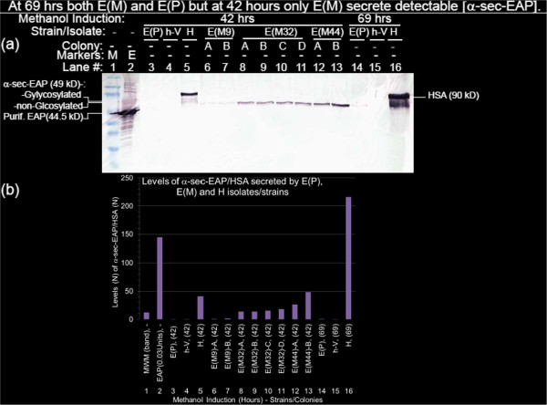
Background: To isolate over-secretors, we subjected to saturation mutagenesis, a strain of P.pastoris exporting E. coli alkaline phosphatase (EAP) fused to the secretory domain of the yeast α factor pheromone through cellular PHO1/KEX2 secretory processing signals as the α-sec-EAP reporter protein. Direct chromogenic staining for α-sec-EAP activity is non-specific as its NBT/BCIP substrate cross-reacts with cellular phosphatases which can be inhibited with Levulinic acid. However, the parental E(P) strain only exports detectable levels of α-sec-EAP at 69 hours and not within the 36 hour period post-seeding required for effective screening with the consequent absence of a reference for secretion. We substituted the endogenous cellular phosphatase activity as a comparative reference for secretion rate and levels as well as for colony alignment while elevating specificity and sensitivity of detection of the exported protein with other innovative modifications of the immuno-chromogenic staining application for screening protein export mutants.

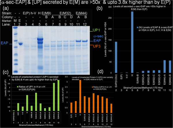
Results: Raising the specificity and utility of staining for α-sec-EAP activity required 5 modifications including some to published methods. These included, exploitation of endogenous phosphatase activity, reduction of the cell/protein burden, establishment of the direct relation between concentrations of transcriptional inducer and exported membrane immobilized protein and concentrations of protein exported into growth media, amplification of immuno-specificity and sensitivity of detection of α-sec-EAP reporter enzyme signal and restriction of staining to optimal concentrations of antisera and time periods. The resultant immuno-chromogenic screen allows for the detection of early secretion and as little as 1.3 fold over-secretion of α-sec-EAP reporter protein by E(M) mutants in the presence of 10 fold -216 fold higher concentrations of HSA.

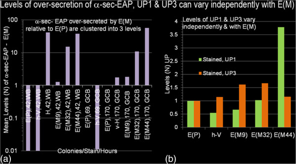
Conclusions: The modified immuno-chromogenic screen is sensitive, specific and has led to the isolation of mutants E(M) over-secreting the α-sec-EAP reporter protein by a minimum of 50 fold higher levels than that exported by non-mutagenized E(P) parental strains. Unselected proteins were also over-secreted.


Figures

Figure 1
Figure 1
Immuno-chromogenic but not chromogenic (direct) stains specifically detect exported proteins. (Figure 1a) Chromogenic stain: Colonies derived from P.pastoris GS115 parental host strains (h) (patches 8, 9) and either strain GS115-pPIC9K Vector (h-V) (patches 12, 13) or exporting Human Serum Albumin, strain GS115-HSAsec (H) (patches 1, 2, 5, 6, 7) or parental strain GS115-EAP(EA3#27) (E(P) exporting α-sec-EAP (patches 3, 4) or the non-exporting strain GS115-β-Gal retaining intracellular β-galactosidase (β-G) (patches 10, 11) were patched onto BMGYA plates overlain with a nitrocellulose (NC) membrane. After 16 hours at 300C the membrane was lifted onto a BMMYA plate and incubated for full induction at 30°C for an additional 24 hours. The NC membrane was then lifted onto Whatman 3MM Chromatography paper soaked with NBT/BCIP and monitored for differential staining of the strains. (Figure 1b) Strains H, α-sec-EAP non-expressor (translational stops in the sequence-E(D2), E(F2), α-sec-EAP expressor E(P), h-V, β-G and h were patched in replicate sets as labeled, onto the top and bottom rows of the top (Figure 1b1, b2, b3) and bottom (Figure b1’, b2’, b3’) halves of a BMGYA plate. This plate was sequentially replicated onto BMMYA plates overlain with nitrocellulose membranes (BMMYA-NC) and supplemented with (0%, 0.01%, 0.025%, 0.05%, 0.075%, 0.1%, 0.2%, 0.3%, 0.5%, 1.0% methanol), MDA plates and BMGYA plates. Representative methanol induction results of series of concentrations are shown in (1b1), (1b1/) at 0.01% (1b2), (1b2/) at 0.05% and (1b3), (1b3/) at 0.2%. Upper and lower halves of nitrocellulose membrane from each plate were respectively immuno-chromogenically stained by reaction with antibodies for HSA with primary/(1°)anti - HSA/(2°)anti-Rb-IgG-AP and for EAP with primary (10)anti-EAP/secondary (2°)anti-Rb-IgG-AP followed by reaction with NBT/BCIP substrate which also serves as substrate for endogenous phosphatases in simultaneous direct chromogenic staining as control for specificity.
Figure 2
Figure 2
Fidelity and selection of replica plated H, E(P) and E(M) colonies secreting/over-secreting HSA and EAP as detected by the immuno-chromogenic assay. (Figure 2a) Concordance of colonies on inductive, selective and rich media. H (400 colonies/88 mm plate) were seeded on BMGYA plates (30°C, 48 hours), sequentially replicated onto (Figure 2a1) BMMYA-NC, (2a2) MDA and (2a3) BMGYA plates and incubated (30°C, 24 hours). (Figure 2a) The filter was lifted and immuno-chromogenically stained for exported HSA. Numbered white arrows (Figure 2a2, 2a3: 2–6) and open white boxes (Figure 2a2, 2a3: box 1) are matched to black arrows (Figure 2a1: 2–6) and box (Figure 2a1: box 1). (b) Selection of α-sec-EAP over-secretor mutants is independent of colony size and seeded strain on nitrocellulose membranes. Primary Screens (Figure 2b1) 400 colony forming units of mutagenized E(P) cells were seeded per 88 mm BMGYA plate and incubated (30°C, 24–36 hours). Colonies were sequentially replicated onto BMMYA-NC plates and BMGYA plates and incubated (30°C, 24 hours and 4°C, overnight). Membrane immobilized proteins were immuno-chromogenically stained for α-sec-EAP activity. Secondary Screens (Figure 2b2): Over-secretor candidates from the primary screen were identified by their intense immuno-chromogenic stain above host cell phosphatase as reference for α-sec-EAP activity. Seventy-three candidate colonies from the primary screen and non-mutagenized E(P) colonies as controls, streaked onto BMGYA plates placed on a 48 square grid and incubated (30°C, 20 hours). Colonies were replicated onto BMMYA-NC, MDA and BMGYA plates and incubated (30°C for 48, 24 and 24 hours, respectively). Immuno-chromogenicaly stained membranes, from BMMYA-NC plates confirmed elevated EAP staining by several of the candidate over-secretors relative to the P(E) strain. Three of these mutant E(M) candidates #9, #M32, #44 (Figure 2b2), were re-purified by streaking onto BMGYA plates, then sequentially replicated onto BMMYA-NC, MDA, and BMGYA followed by immuno-chromogenic staining. Purified single candidate colonies (2–4 replicates) (Figure 2b3), #9, #32 and #44) were inoculated for confirmation as over-secretors by Western Blot and Direct Staining with Gel Code Blue (GCB) staining of electrophoretically resolved proteins.
Figure 3
Figure 3
At 69 hrs both E(M) and E(P) but at 42 hours only E(M) secrete detectable [α-sec-EAP]. Culture supernatants of, E(P), h-V and H were harvested after methanol induction for 42 hrs (Figure 3a, lanes 3, 4 and 5) and 69 hours (Figure 3a, lanes 14, 15 and 16), 2–4 colonies per candidate E(M) mutants #9, #32 and #44 (from the re-streaked tertiary (3°) screen e.g. in Figure 2b3) after 42 hours of methanol induction (lanes 6 to 13) were electrophoresed, proteins transferred to NC membranes, sequentially annealed with primary (1°)anti-HSA/primary (1°)anti-EAP) and secondary (2°) anti-Rb-IgG-AP antibodies, washed, reacted with NBT/BCIP substrate and AP terminator as described in the materials and methods section. (Figure 3b). Levels of α-sec-EAP/HSA secreted by E(P), E(M) and H isolates/strains. The filter was scanned and saved as a TIFF file. Pixel densities of the proteins in the regions of interest (ROI) corresponding to, EAP (0.03 U) and MWM standards (Figure 3b, lanes 1, 2), α-sec-EAP (Figure 3b, lanes 3, 4, 6–15) and HSA (Figure 3b, lanes 5 and 16) in the Western Blot shown in Figure 3(a) were quantified with the NIH Image J Densitometric software. The respective pixel densities were normalized to E(P) at 69 Hrs (Figure 3b, lane 14) and plotted with Excel software. Based on this quantification the levels of secretion of α-sec-EAP confirmed the 3 different classes of E(M) mutants visually identified in the initial colony isolation as E(M9)-A, E(M9)-B (Figure 3b, lanes 6, 7), E(M32)-A, E(M32)-B, E(M32)-C, E(M32)-D (lanes 8–11), E(M44)-A and E(M44)-B (Figure 3b, lanes 12, 13).
Figure 4
Figure 4
Levels of over-secretion of α-sec-EAP, UP1 and UP3 can vary independently with E(M). Values or mean values of ROIs corresponding to α-sec-EAP and HSA (42 and 69 hours inductions) from Western Blot Analysis normalized to E(P) 69 hours (Figure 3) and Gel Code Blue stained gels from 170 hour inductions normalized to UP3 from E(P) (Figure 5) were plotted as semi-log scales (Figure 4a, graphs WB and GCB). Plotted values were taken from tables 2, 3 and 4. However, to permit graphical resolution of low values, results of HSA secreted after 69 hours and 170 hours of induction were omitted from the plots. Mean values of UP1 and UP3 expressed as their ratios in E(P)/E(P), v-H/E(P) and all E(M)/E(P) were plotted (Figure 4b).
Figure 5
Figure 5
[α-sec-EAP] and [unselected proteins,UP] secreted by E(M) are >50 and upto 4 fold higher than those secreted by E(P). (Figure 5a) Incubation of the panel of cultures, shown in Figure 3 (with the exception of E(M9)-B, in lane 6) was for 170 hours with supplementation of methanol inducer at 24 hour intervals. Culture supernatants were harvested as determine in the materials and methods. Aliquots (20 μL) of cultures of E(P), h-V, H, E(M9)-B, EM32)-A,-B,-C,-D and E(M44)-A,-B were loaded in the order shown above the lanes (lanes 3–12) and co-electrophoresed with broad range markers (lane 1) and EAP purified from E.coli (0.08 units/lane, lanes2) through denaturing (4%-20%) polyacrylamide gradient gels with Laemmli electrophoresis buffer. The gel was stained with Gel Code Blue (GCB) scanned and saved as TIFF files. The respective ROIs of proteins corresponding to EAP, α-sec-EAP, unselected protein-1 (UP1) and unselected protein 3 (UP3) are identified and color coded on the left and right of the gel. The ROIs corresponding to the above 4 proteins were densitometrically scanned and normalized to UP3 in lane 3 and plotted as their respective ratios to E(P). Normalized ROIs corresponding to EAP, α-sec-EAP and HSA in E(P), v-H, H and E(M) were plotted with Excel programs (Figure 5b). Normalized ROIs corresponding to UP1 and UP3 were plotted as their ratios in EAP/E(P), E(P)/E(P), v-H/E(P), H/E(P) and all E(M)/E(P) (Figure 5c, 5d).

Similar articles

Cited by

References

    1. Koutz P, Davis GR, Stillman C, Barringer K, Cregg J, Thill G. Structural comparison of the Pichia pastoris Alcohol Oxidase Genes. Yeast. 1989;5:167–177. doi: 10.1002/yea.320050306. - DOI - PubMed
    1. Cregg JM, Cereghino JL, Shi J, Higgins DR. Recombinant protein expression in Pichia Pastoris. Mol Biotechnol. 2006;16:23–52. - PubMed
    1. Cereghino GP, Cereghino JL, Ilgen C, Cregg JM. Production of recombinant proteins in fermenter cultures of the yeast, Pichia pastoris. Curr Opin Biotechnol. 2002;13(4):329–332. doi: 10.1016/S0958-1669(02)00330-0. - DOI - PubMed
    1. Gelissen G, Kunze G, Gaillardin C, Cregg JM, Berardi E, Veenhuis M, vander Kei I. New yeast expression platforms based on methylotrophic Hanensula polymorpha and Pichia pastoris and on dimorphic Arxula adeninivorus and Yarowia lipolytica - a comparison. FEMS Yeast Res. 2005;5(11):1079–1096. doi: 10.1016/j.femsyr.2005.06.004. - DOI - PubMed
    1. Julien C. Production of Humanlike Recombinant Proteins in Pichia pastoris. From Expression Vectors to Fermentation Strategy. Bioprocess Int. 2006;4(1):22–31.

LinkOut - more resources