Review
. 2013 Apr;3(2):137-42.
doi: 10.1016/j.coviro.2013.03.009. Epub 2013 Apr 18.

Viral precursor polyproteins: keys of regulation from replication to maturation

Affiliations
Review

Viral precursor polyproteins: keys of regulation from replication to maturation

Samantha A Yost et al. Curr Opin Virol. 2013 Apr.

Abstract

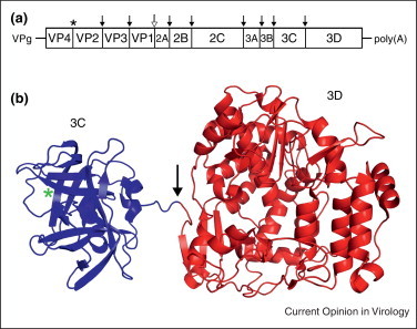
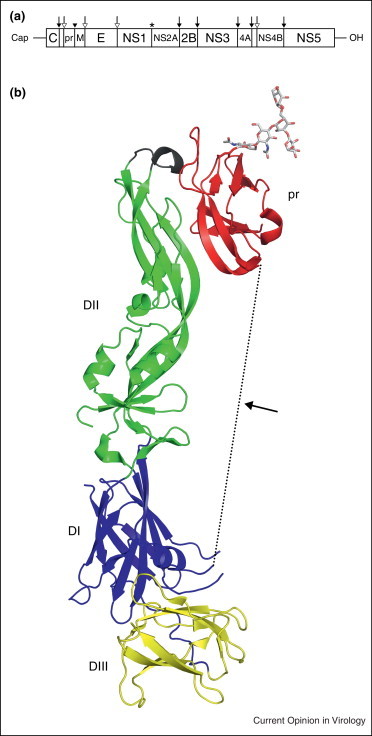
Many viruses use a replication strategy involving the translation of a large polyprotein, which is cleaved by viral and/or cellular proteases. Several of these viruses severely impact human health around the globe, including HIV, HCV, Dengue virus, and West Nile virus. This method of genome organization has many benefits to the virus such as condensation of genetic material, as well as temporal and spatial regulation of protein activity depending on polyprotein cleavage state. The study of polyprotein precursors is necessary to fully understand viral infection, and identify possible new drug targets; however, few atomic structures are currently available. Presented here are structures of four recent polyprotein precursors from viruses with a positive sense RNA genome.


Figures

Figure 1
Figure 1
Alphavirus genome organization and P23 structure. (a) RNA genome organization of Sindbis virus with 5′ cap and 3′ poly(A) tail. Polyprotein cleavage sites are denoted with an asterisk (nsP2 protease), solid black arrow (Sindbis virus capsid autoprotease), solid black arrowheads (cellular signalase), and hollow arrow (cellular furin). (b) P23 structure (PDB ID: 4GUA) highlighting the nsP2 protease (blue) and methyltransferase-like (cyan) domains and nsP3 macro (yellow) and zinc binding (red) domains. The protease active site is denoted with a green asterisk, zinc ion with a gray sphere, and the P2/3 cleavage site with an arrow. (c) nsP3 macro and zinc binding domains of the P23 structure shown in ribbon format, with coloring similar to panel B. The extended linker between the two nsP3 domains creates a ring-like structure which is fitted around nsP2.
Figure 2
Figure 2
Organization of poliovirus genome and 3CD structure. (a) The RNA genome organization of poliovirus with 5′ covalently linked viral protein VPg and 3′ poly(A) tail. Polyprotein cleavage sites are denoted by an asterisk (autoprotease), solid black arrow (3C or 3CD protease), and a hollow arrow (2A protease). (b) Ribbons representation of the 3CD structure (PDB ID: 2IJD) with a green asterisk indicating the 3C protease active site and an arrow showing the cleavage site.
Figure 3
Figure 3
Genomic organization of Flavivirus genome and the structure of prM-E. (a) The genome organization of flavivirus shown with a 5′ cap and 3′ OH. Polyprotein cleavage sites are denoted by an asterisk (unknown protease), solid black arrow (NS2B-NS3 protease), solid black arrowhead (cellular furin), and a hollow arrow (cellular signalase). (b) Structure of prM-E (PDB ID: 3C5X) highlighting pr (red) with E domains DI (blue), DII (green), and DIII (yellow). The fusion loop of E protein is shown in black. The M protein and linker region, which are missing in the structure, are represented as a dotted line.
Figure 4
Figure 4
HIV Gag polyprotein organization and structure. (a) Schematic representation of the domain organization of HIV-1 Gag polyprotein. Cleavage sites by viral PR protein are denoted with a solid, black arrow. (b) N-terminal 283 residues of the Gag polyprotein (PDB ID: 1L6N) including MA (red) and the N-terminal portion of CA (blue). The post cleavage form of CA (PDB ID: 1GWP) is shown in gray with a box highlighting the N-terminal β hairpin formed after protease digestion. The cleavage site is noted with an arrow.

References

    1. Andres G., Alejo A., Salas J., Salas M.L. African swine fever virus polyproteins pp220 and pp62 assemble into the core shell. J Virol. 2002;76:12473–12482. - PMC - PubMed
    1. Griffin D.E. Alphaviruses. In: David M., Knipe P.M.H., editors. vol 1. Lippincott Williams & Wilkins; 2007. pp. 1023–1067. (Fields Virology).
    1. de Groot R.J., Hardy W.R., Shirako Y., Strauss J.H. Cleavage-site preferences of Sindbis virus polyproteins containing the non-structural proteinase. Evidence for temporal regulation of polyprotein processing in vivo. EMBO J. 1990;9:2631–2638. - PMC - PubMed
    1. Vasiljeva L., Merits A., Golubtsov A., Sizemskaja V., Kääriäinen L., Ahola T. Regulation of the sequential processing of Semliki Forest virus replicase polyprotein. J Biol Chem. 2003;278:41636–41645. - PubMed
    1. Shirako Y., Strauss J.H. Regulation of Sindbis virus RNA replication: uncleaved P123 and nsP4 function in minus-strand RNA synthesis, whereas cleaved products from P123 are required for efficient plus-strand RNA synthesis. J Virol. 1994;68:1874–1885. - PMC - PubMed

Publication types

LinkOut - more resources