Review
. 2013 Apr;3(2):103-10.
doi: 10.1016/j.coviro.2013.03.008. Epub 2013 Apr 18.

The polymerase of negative-stranded RNA viruses

Affiliations
Review

The polymerase of negative-stranded RNA viruses

Benjamin Morin et al. Curr Opin Virol. 2013 Apr.

Abstract

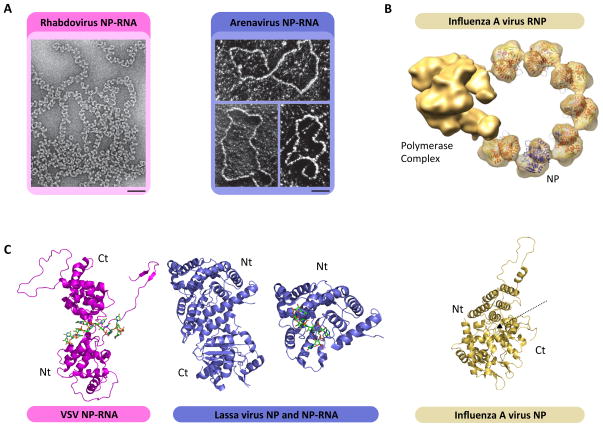
Negative-sense (NS) RNA viruses deliver into cells a mega-dalton RNA-protein complex competent for transcription. Within this complex, the RNA is protected in a nucleocapsid protein (NP) sheath which the viral polymerase negotiates during RNA synthesis. The NP-RNA templates come as nonsegmented (NNS) or segmented (SNS), necessitating distinct strategies for transcription by their polymerases. Atomic-level understanding of the NP-RNA of both NNS and SNS RNA viruses show that the RNA must be transiently dissociated from NP during RNA synthesis. Here we summarize and compare the polymerases of NNS and SNS RNA viruses, and the current structural data on the polymerases. Those comparisons inform us on the evolution of related RNA synthesis machines which use two distinct mechanisms for mRNA cap formation.


Figures

Figure 1
Figure 1. Structural architecture and organization of NS RNA viral polymerases
Conserved architecture and domain organization found in nonsegmented (top, purple) and segmented (bottom, orange) polymerases. The linear amino acid sequence of L and the tripartite influenza virus polymerase contain highly conserved regions dedicated to RNA synthesis (blue boxes). L and the influenza virus polymerase also contain blocks of conservation dedicated to 5′ cap formation (maroon boxes), including an endonuclease domain for cap-snatching (domain I or PA, segmented NS RNA viruses) or PRNTase / MTase domains for de novo cap synthesis (domains V and VI, nonsegmented NS RNA viruses). The regions containing cap formation enzymatic activities are also required for RNA synthesis, and the exact function of domains II and IV in the L protein of segmented NS RNA viruses remains unknown. EM of purified L from Machupo virus and vesicular stomatitis virus reveals a shared structural architecture conserved within NS RNA viral L proteins. L consists of a central ring-like RNA polymerase domain (blue highlight in cartoon) and a large appendage dedicated to 5′ cap formation (maroon highlight in cartoon) attached with a flexible linkage. As described in the text, comparison with EM analysis and 3D reconstruction of the influenza virus polymerase complex provides further insight into the architecture of L[19,22,27]. (Fig. adapted from ref. 4)
Figure 2
Figure 2. Architecture of the NP-RNA complex
(A) EM images of rhabdovirus (NNS RNA viruses, magenta) and arenavirus (SNS RNA viruses, blue) NP-RNA genomic template isolated from purified virions. (Scale bar: 50 nm) [37,52]. (B) Pseudoatomic model of influenza A virus RNP. The tripartite polymerase complex is bound to a truncated NP-RNA template. Atomic structures of the NP are docked inside the EM density. Adapted from (ref). (C) Crystal structure of VSV (magenta), Lassa virus (blue) and influenza A virus NP. The N-terminal (Nt) and C-terminal (Ct) domains are indicated. VSV NP is shown in complex with a RNA oligomer that binds NP in between the N and C-teminal domain of the protein. The full Lassa NP is represented devoid of RNA (left), while the N-terminal domain is shown in complex with a RNA oligomer (right). The influenza A NP is represented free of RNA, the putative RNA binding site is indicated by a dashed arrow.
Figure 3
Figure 3. Molecular architecture of VSV P and L
(A) Schematic organization of VSV P. P consists of three domains connected by two intrinsically disordered regions (Black line). The N-terminal (Nt) and C-terminal (Ct) are indicated. The P N-terminal domain (PNTD, red) contains the RNA-free N (N0) binding region, the central domain (PCD, orange) consists of an oligomerization region and the C-terminal domain (PCTD, green) is involved in the N-RNA binding. (B) Crystallographic structure of VSV PNTD (top), a dimer of PCD (middle) and PCTD (bottom). (C) Structure of PNTD in complex with N0 (magenta) (top) and PCTD in complex with the C-teminal domain of N (magenta) (bottom). (D) EM image (left) and model (right) of L organization. L is arranged with a core ring structure harboring the RdRP domain and a flexible appendage containing the activities necessary for cap formation (capping + methylation). The arrows depict putative flexible linkers. (E) EM image (right) and model (left) of the L-P complex. Upon binding to P or P41-106, the appendage of L undergoes a structural rearrangement. When L binds full length P, the L-P complex is isolated in two forms a monomer and a dimer in which the L pairs are likely bridged by interaction with an oligomer of P (top). The arrow represents the variable orientation of L proteins in the dimers. When L binds P41-106 the L-P41-106 complex exits only as a monomer (bottom).
Figure 4
Figure 4. VSV L in vitro RNA synthesis assay
(A) De novo RNA synthesis by VSV L, in the absence and presence of P, on a synthetic template corresponding to the first 19 nt of the leader (Le19). Activity assays were set using 0.2 μM of Le19, 0.2 μM of protein and [α32P]-GTP as the radio-labeled nucleotide. Reactions were quenched by the addition EDTA/formamide and analyzed on a 20% polyacrylamide/7 M urea gel. De novo initiation product sizes are indicated on the left. (B) RNA synthesis by VSV L on a naked 50 (Le50) nucleotides long RNAs, and the encapsidated RNA (N-RNA) templates. (C) RNA synthesis by MACV L on a naked RNA (left), or a naked mutated RNA (right).

References

    1. Geisbert TW, Jahrling PB. Exotic emerging viral diseases: progress and challenges. Nat Med. 2004;10:S110–121. - PubMed
    1. Pappu HR, Jones RA, Jain RK. Global status of tospovirus epidemics in diverse cropping systems: successes achieved and challenges ahead. Virus Res. 2009;141:219–236. - PubMed
    1. Whelan SP, Barr JN, Wertz GW. Transcription and replication of nonsegmented negative-strand RNA viruses. Curr Top Microbiol Immunol. 2004;283:61–119. - PubMed
    1. Kranzusch PJ, Whelan SP. Architecture and regulation of negative-strand viral enzymatic machinery. RNA Biol. 2012;9:941–948. - PMC - PubMed
    1. Morin B, Whelan SP. La protéine L des Mononegavirales. Virologie. 2012;16:258–268. - PubMed

Publication types

MeSH terms

LinkOut - more resources