

Diagnosis of relevant prostate cancer using supplementary cores from magnetic resonance imaging-prompted areas following multiple failed biopsies
- PMID: 23602725
- PMCID: PMC3676721
- DOI: 10.1016/j.mri.2013.02.007
Diagnosis of relevant prostate cancer using supplementary cores from magnetic resonance imaging-prompted areas following multiple failed biopsies
Abstract
Objectives: To establish the value of MRI in targeting re-biopsy for undiagnosed prostate cancer despite multiple negative biopsies and determine clinical relevance of detected tumors.
Materials and methods: Thirty-eight patients who underwent MRI after 2 or more negative biopsies due to continued clinical suspicion and later underwent TRUS-guided biopsy supplemented by biopsy of suspicious areas depicted by MRI were identified. Diagnostic performance of endorectal 3T MRI in diagnosing missed cancer foci was assessed using biopsy results as the standard of reference. Ratio of positive biopsies using systematic versus MRI-prompted approaches was compared. Gleason scores of detected cancers were used as surrogate for clinical relevance.
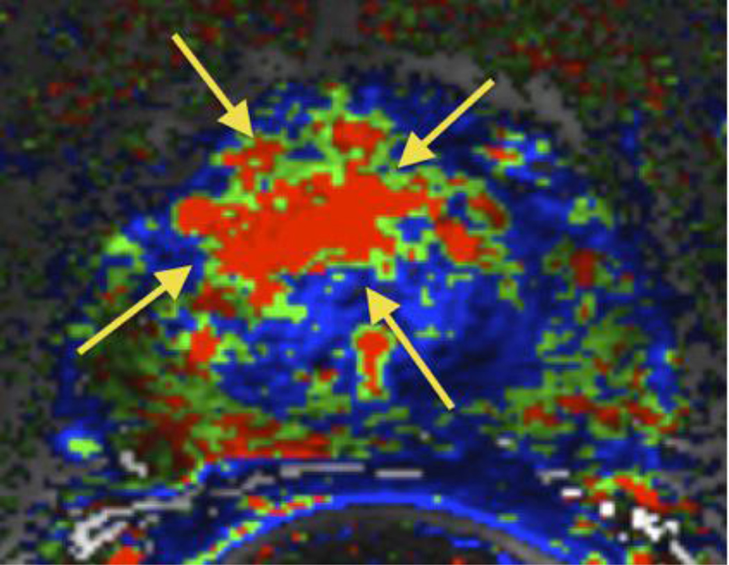
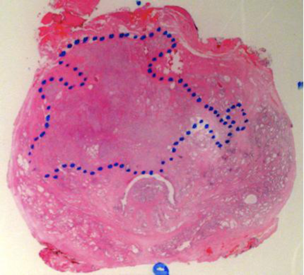
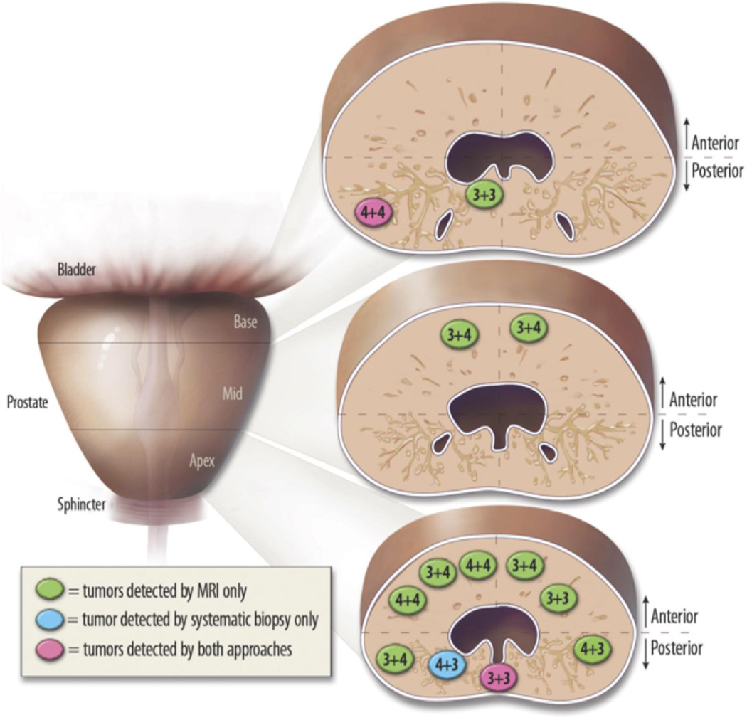
Results: Thirty-four percent of patients who underwent MRI before re-biopsy had prostate cancer on subsequent biopsy. The positive biopsy yield with systematic sampling was 23% versus 92% with MRI-prompted biopsies(p<0.0001). Seventy-seven percent of tumors were detected exclusively in the MRI-prompted zones. Sensitivity, specificity, positive predictive value, negative predictive value and accuracy of MRI to provide a positive biopsy were 92%, 60%, 55%, 94% and 71%, respectively. The anterior gland and apical regions contained most tumors; 75% of cancers detected by MRI-prompted biopsy had Gleason score≥7.
Conclusions: Clinically relevant tumors missed by multiple TRUS-guided biopsies can be detected by a MRI-prompted approach.
Copyright © 2013 Elsevier Inc. All rights reserved.
Figures

References
-
- Durkan GC, Greene DR. Elevated serum prostate specific antigen levels in conjunction with an initial prostatic biopsy negative for carcinoma :who should undergo a repeat biopsy? BJU Int. 1999;83(1):34–38. - PubMed
-
- Djavan B, et al. Repeat prostate biopsy: who how and when? A review. Eur Urol. 2002;42(2):93–103. - PubMed
-
- Scattoni V, et al. Extended and saturation prostatic biopsy in the diagnosis and characterisation of prostate cancer: a critical analysis of the literature. European urology. 2007;52(5):1309–1322. - PubMed
-
- Ashley RA, et al. Reassessing the diagnostic yield of saturation biopsy of the prostate. European urology. 2008;53(5):976–981. - PubMed
-
- Lawrentschuk N, et al. 'Prostatic evasive anterior tumours': the role of magnetic resonance imaging. BJU Int. 2009;105(9):1231–1236. - PubMed
Publication types
MeSH terms
Grants and funding
LinkOut - more resources
Full Text Sources
Other Literature Sources
Medical
