. 2013 Jun;182(6):2310-21.
doi: 10.1016/j.ajpath.2013.02.037. Epub 2013 Apr 17.

Inflammatory cytokines associated with degenerative disc disease control aggrecanase-1 (ADAMTS-4) expression in nucleus pulposus cells through MAPK and NF-κB

Affiliations

Inflammatory cytokines associated with degenerative disc disease control aggrecanase-1 (ADAMTS-4) expression in nucleus pulposus cells through MAPK and NF-κB

Ye Tian et al. Am J Pathol. 2013 Jun.

Abstract

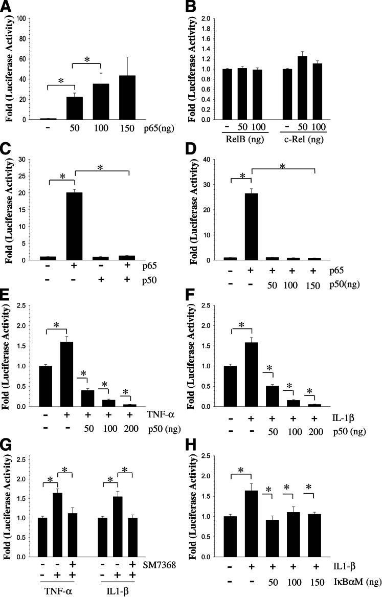
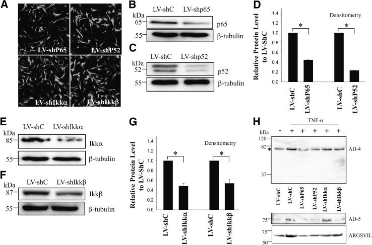
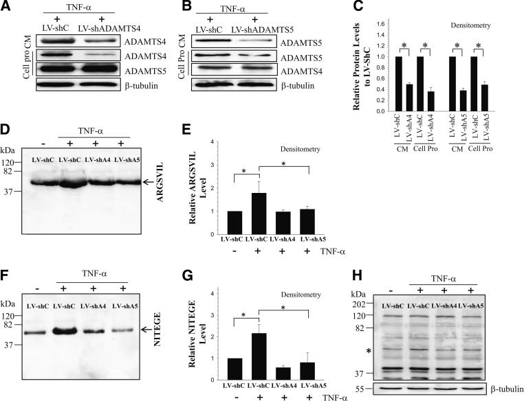
We investigated TNF-α and IL-1β regulation of ADAMTS-4 expression in nucleus pulposus (NP) cells and its role in aggrecan degradation. Real-time quantitative RT-PCR, Western blotting, and transient transfections with rat NP cells and lentiviral silencing with human NP cells were performed to determine the roles of MAPK and NF-κB in cytokine-mediated ADAMTS-4 expression and function. ADAMTS4 expression and promoter activity increased in NP cells after TNF-α and IL-1β treatment. Treatment of cells with MAPK and NF-κB inhibitors abolished the inductive effect of the cytokines on ADAMTS4 mRNA and protein expression. Although ERK1, p38α, p38β2, and p38γ were involved in induction, ERK2 and p38δ played no role in TNF-α-dependent promoter activity. The inductive effect of p65 on ADAMTS4 promoter was confirmed through gain and loss-of-function studies. Cotransfection of p50 completely blocked p65-mediated induction. Lentiviral transduction with shRNA plasmids shp65, shp52, shIKK-α, and shIKK-β significantly decreased TNF-α-dependent increase in ADAMTS-4 and -5 levels and aggrecan degradation. Silencing of either ADAMTS-4 or -5 resulted in reduction in TNF-α-dependent aggrecan degradation in NP cells. By controlling activation of MAPK and NF-κB signaling, TNF-α and IL-1β modulate expression of ADAMTS-4 in NP cells. To our knowledge, this is the first study to show nonredundant contribution of both ADAMTS-4 and ADAMTS-5 to aggrecan degradation in human NP cells in vitro.


Figures

Figure 1
Figure 1
Expression and cytokine dependency of ADAMTS-4 in rat NP cells. A: RT-qPCR analysis shows ADAMTS-4 was expressed at a very low level in adult rat NP and annulus fibrosus (AF) tissues. B: Western blot analysis of ADAMTS4 expression in adult disk tissues reveals bands at 58 and 73 kDa, representing mature/processed protein. C and D: RT-qPCR analysis of ADAMTS4 expression by rat NP cells treated with the cytokines TNF-α (C) and IL-1β (D) for 24 hours. There was a dose-dependent increase in ADAMTS4 mRNA expression by the cytokine treatment. E and F: Western blot analysis of NP cells indicates increased expression of ADAMTS-4 after TNF-α and IL-1β treatment. G: Schematic of ADAMTS4 promoter constructs, showing important transcription factor and regulatory elements. H: Treatment of NP cells with TNF-α and IL-1β resulted in significant induction of ADAMTS4 promoter activity. Data are expressed as means ± SEM from three independent experiments. P < 0.05. Ctrl, control.
Figure 2
Figure 2
Modulation of cytokine-dependent expression of ADAMTS-4 and -5 expression by NF-κB and MAPK signaling in rat NP cells. A and B: Western blot analysis of NF-κB and MAPK signaling proteins after treatment of NP cells with TNF-α (A) and IL-1β (B). Cytokine treatment induced phosphorylation of p65, p38, ERK, JNK within the first 15 minutes. No appreciable change was observed in expression of p65, p38, ERK, and JNK. C–F: RT-qPCR analysis of ADAMTS4 and ADAMTS5 expression by NP cells after treatment with TNF-α (C and E) or IL-1β (D and F) for 24 hours with or without inhibitors for NF-κB (SM7368, 10 μmol/L), p38 (SB203580, 10 μmol/L), ERK (PD98059, 10 μmol/L), and JNK (SP600125, 10 μmol/L). Inhibition of NF-κB signaling and MAPK signaling resulted in a significant blocking of cytokine-dependent induction in ADAMTS4 and ADAMTS5 mRNA expression. G and H: Western blot analysis indicates that treatment with NF-κB and MAPK inhibitors completely abolished ADAMTS-4 protein induction (the bands are indicated by asterisk) by TNF-α (G) and IL-1β (H). Data are expressed as means ± SEM from three independent experiments. P < 0.05.
Figure 3
Figure 3
MAPK signaling controls ADAMTS4 promoter activity in rat NP cells. A and B: Cotransfection of rat NP cells with DN-p38α abolished TNF-α–dependent (A) and IL-1β–dependent (B) induction in ADAMTS4 promoter activity. C and D: IL-1β–dependent increase in ADAMTS4 promoter activity was blocked by DN-p38β2 (C) and DN-p38γ (D). E and F: DN-ERK1 suppressed induction in ADAMTS4 promoter activity by TNF-α (C) and IL-1β (D) treatment. For IL-1β treatment, inhibition was seen only at the highest dose (150 ng). G: NP cells were cotransfected with LIP and ADAMTS4 promoter, and luciferase activity was measured after TNF-α treatment. Addition of LIP further increased TNF-α–dependent ADAMTS-4 reporter activity. H:ADAMTS4 promoter activity was measured after cotransfection with LAP2. LAP2 suppressed ADAMTS4 promoter activity. Data are expressed as means ± SEM from three independent experiments. P < 0.05.
Figure 4
Figure 4
NF-κB regulation of ADAMTS-4 expression. A: Rat NP cells were transfected with RelA/p65, and ADAMTS4 promoter activity was measured. There was a dose-dependent increase in promoter activity up to 100 ng of p65. B: Cotransfection with RelB and c-Rel had no effect on ADAMTS4 promoter activity in rat NP cells. C: Rat NP cells were cotransfected with RelA/p65 and/or p50, and promoter activity was measured. When p65 and p50 were added together, p50 significantly blocked p65-mediated induction in promoter activity. D: Rat NP cells were cotransfected with p65 alone and with increasing doses of p50. Even at 50 ng, p50 completely blocked p65-mediated induction of promoter activity. E and F: Rat NP cells cotransfected with p50, and ADAMTS4 promoter activity was measured after TNF-α (E) and IL-1β (F) treatment. Cytokine-mediated induction in promoter activity was completely blocked by p50. G: TNF-α– and IL-1β–mediated induction in promoter activity was completely blocked by the NF-κB inhibitor SM7368. H: Cotransfection of cells with DN-NF-κB/IκBαM resulted in a significant inhibition of IL-1β–dependent ADAMTS4 promoter activity. Data are expressed as means ± SEM from three independent experiments. P < 0.05.
Figure 5
Figure 5
Regulation of ADAMTS-4 and -5 expression by NF-κB. A: Immunofluorescence detection of YFP in human NP cells transduced with lentivirus coexpressing YFP and NF-κB pathway–specific shRNAs (LV-shp65, LV-shp52, LV-shIKKα, LV-shIKKβ) show high transduction efficiency. B–G: Western blot analysis of cells transduced with control lentivirus LV-shC and LV-shp65 (B), LV-shp52 (C), LV-shIKKα (E), and LV-shIKKβ (F). Expression of p65, p52, IKK-α, and IKK-β was suppressed by corresponding shRNAs, compared with cells transduced with a lentivirus expressing control shRNA. Densitometric analysis of p65 and p52 in cells transduced with LV-shp65 and LV-shp52 (D) and of IKK-α and IKK-β in cells transduced with LV-shIKKα and LV-shIKKβ (G). H: Western blot analysis of ADAMTS-4 (AD-4), ADAMTS-5 (AD-5), and aggrecan neoepitope (ARGSVIL) in human NP cells infected with LV-shC and LV-shp65, LV-shp52, LV-shIKKα, and LV-shIKKβ after TNF-α treatment. Note that the TNF-α–dependent increase in ADAMTS-4 (the band is indicated by an asterisk), ADAMTS-5, and aggrecan neoepitope (ARGSVIL) levels is significantly blocked by suppression of components of the NF-κB signaling pathway. Data are expressed as means ± SEM from three independent experiments. P < 0.05. Original magnification, ×20.
Figure 6
Figure 6
ADAMTS-4 and ADAMTS-5 promote aggrecan degradation in human NP cells. A and B: Western blot analysis of human NP cells infected with control lentivirus (LV-shC) and lentivirus expressing shRNA ADAMTS-4 (LV-shADAMTS4) (A) and shRNA ADAMTS-5 (LV-shADAMTS5) (B) plasmids. Compared with control cells, expression of ADAMTS-4 and ADAMTS-5 was suppressed by shRNA shADAMTS-4 and shADAMTS-5 in both the conditioned medium and cell protein. C: Densitometric analysis of multiple blots from the experiment presented in panels A and B. D and E: Western blot (D) and corresponding densitometric analysis (E) of aggrecan neoepitope (ARGSVIL) in conditioned medium of cells treated with TNF-α. The level of ARGSVIL was significantly reduced after cytokine treatment in cells transduced with LV-shADAMTS-4 (LV-shA4) and LV-shADAMTS-4 (LV-shA5), compared with control (LV-shC). F and G: Western blot (F) and corresponding densitometric analysis (G) of aggrecan neoepitope (NITEGE) in concentrated conditioned medium of cells treated with TNF-α. The level of NITEGE was significantly reduced after TNF-α treatment in ADAMTS-4 and ADAMTS-5 silenced NP cells. H: Western blot analysis of NITEGE in cell-associated protein (the band is indicated by an asterisk) shows a significant reduction in levels after cytokine treatment in ADAMTS-4 and ADAMTS-5 silenced cells, compared with controls. Data are expressed as means ± SEM from three independent experiments. P < 0.05. Cell Pro, cell protein; CM, conditioned medium.

References

    1. Stemple D.L. Structure and function of the notochord: an essential organ for chordate development. Development. 2005;132:2503–2512. - PubMed
    1. Feng H., Danfelter M., Strömqvist B., Heinegård D. Extracellular matrix in disc degeneration. J Bone Joint Surg Am. 2006;88(Suppl 2):25–29. - PubMed
    1. Setton L.A., Chen J. Mechanobiology of the intervertebral disc and relevance to disc degeneration. J Bone Joint Surg Am. 2006;88(Suppl 2):52–57. - PubMed
    1. Ng L., Grodzinsky A.J., Patwari P., Sandy J., Plaas A., Ortiz C. Individual cartilage aggrecan macromolecules and their constituent glycosaminoglycans visualized via atomic force microscopy. J Struct Biol. 2003;143:242–257. - PubMed
    1. Urban J.P., Roberts S. Degeneration of the intervertebral disc. Arthritis Res Ther. 2003;5:120–130. - PMC - PubMed

Publication types

MeSH terms