

Macrophages are essential for the early wound healing response and the formation of a fibrovascular scar
- PMID: 23602833
- PMCID: PMC3668032
- DOI: 10.1016/j.ajpath.2013.02.032
Macrophages are essential for the early wound healing response and the formation of a fibrovascular scar
Abstract
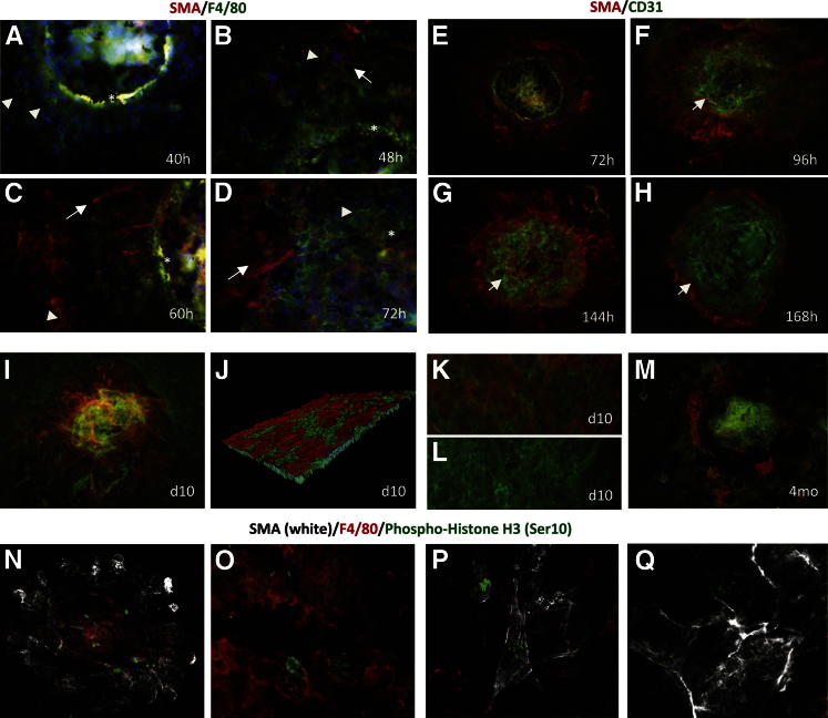
After wounding, multiple cell types interact to form a fibrovascular scar; the formation and cellular origins of these scars are incompletely understood. We used a laser-injury wound model of choroidal neovascularization in the eye to determine the spatiotemporal cellular events that lead to formation of a fibrovascular scar. After laser injury, F4/80(+) myeloid cells infiltrate the wound site and induce smooth muscle actin (SMA) expression in adjacent retinal pigment epithelial cells, with subsequent formation of a SMA(+)NG2(+) myofibroblastic scaffold, into which endothelial cells then infiltrate to form a fibrovascular lesion. Cells of the fibrovascular scaffold express the proangiogenic factor IL-1β strongly, whereas retinal pigment epithelial cells are the main source of VEGF-A. Subsequent choroidal neovascularization is limited to the area demarcated by this myofibroblastic scaffold and occurs independently of epithelial- or myeloid-derived VEGF-A. The SMA(+)NG2(+) myofibroblastic cells, F4/80(+) macrophages, and adjacent epithelial cells actively proliferate in the early phase of the wound healing response. Cell-lineage tracing experiments suggest that the SMA(+)NG2(+) myofibroblastic scaffold originates from choroidal pericyte-like cells. Targeted ablation of macrophages inhibits the formation of this fibrovascular scaffold, and expression analysis reveals that these macrophages are Arg1(+)YM1(+)F4/80(+) alternatively activated M2-like macrophages, which do not require IL-4/STAT6 or IL-10 signaling for their activation. Thus, macrophages are essential for the early wound healing response and the formation of a fibrovascular scar.
Copyright © 2013 American Society for Investigative Pathology. Published by Elsevier Inc. All rights reserved.
Figures


References
-
- Marneros A.G., She H., Zambarakji H., Hashizume H., Connolly E.J., Kim I., Gragoudas E.S., Miller J.W., Olsen B.R. Endogenous endostatin inhibits choroidal neovascularization. FASEB J. 2007;21:3809–3818. - PubMed
-
- Sakurai E., Anand A., Ambati B.K., van Rooijen N., Ambati J. Macrophage depletion inhibits experimental choroidal neovascularization. Invest Ophthalmol Vis Sci. 2003;44:3578–3585. - PubMed
-
- Bohgaki M., Kitaguchi H. Conversion of cultured monocytes/macrophages into endothelial-like cells through direct contact with endothelial cells. Int J Hematol. 2007;86:42–48. - PubMed
Publication types
MeSH terms
Substances
Grants and funding
LinkOut - more resources
Full Text Sources
Other Literature Sources
Medical
Molecular Biology Databases
Research Materials
Miscellaneous
