

Untangling the web: mechanisms underlying ER network formation
- PMID: 23602970
- PMCID: PMC3729797
- DOI: 10.1016/j.bbamcr.2013.04.009
Untangling the web: mechanisms underlying ER network formation
Abstract
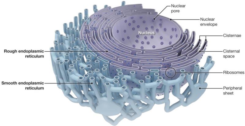
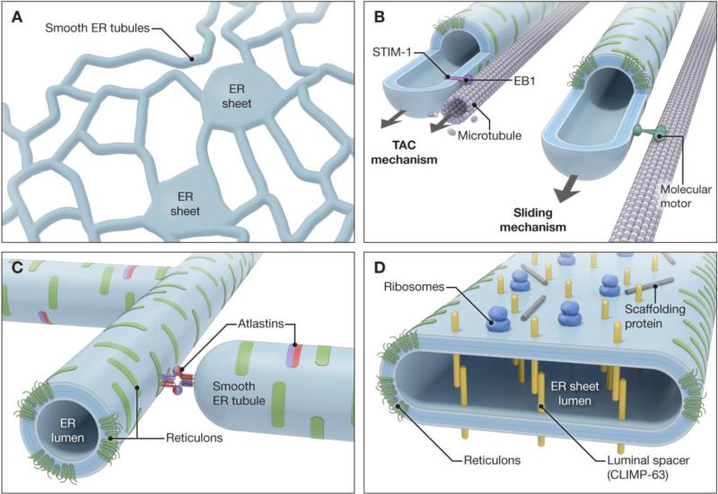
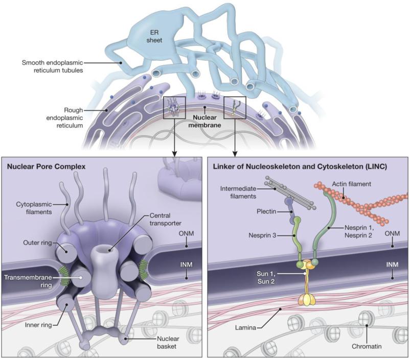
The ER is a continuous membrane system consisting of the nuclear envelope, flat sheets often studded with ribosomes, and a polygonal network of highly-curved tubules extending throughout the cell. Although protein and lipid biosynthesis, protein modification, vesicular transport, Ca(2+)dynamics, and protein quality control have been investigated in great detail, mechanisms that generate the distinctive architecture of the ER have been uncovered only recently. Several protein families including the reticulons and REEPs/DP1/Yop1p harbor hydrophobic hairpin domains that shape high-curvature ER tubules and mediate intramembrane protein interactions. Members of the atlastin/RHD3/Sey1p family of dynamin-related GTPases interact with the ER-shaping proteins and mediate the formation of three-way junctions responsible for the polygonal structure of the tubular ER network, with Lunapark proteins acting antagonistically. Additional classes of tubular ER proteins including some REEPs and the M1 spastin ATPase interact with the microtubule cytoskeleton. Flat ER sheets possess a different complement of proteins such as p180, CLIMP-63 and kinectin implicated in shaping, cisternal stacking and cytoskeletal interactions. The ER is also in constant motion, and numerous signaling pathways as well as interactions among cytoskeletal elements, the plasma membrane, and organelles cooperate to position and shape the ER dynamically. Finally, many proteins involved in shaping the ER network are mutated in the most common forms of hereditary spastic paraplegia, indicating a particular importance for proper ER morphology and distribution in large, highly-polarized cells such as neurons. This article is part of a Special Issue entitled: Functional and structural diversity of endoplasmic reticulum.
Keywords: Atlastin; Endoplasmic reticulum; Hereditary spastic paraplegia; Morphology; REEP; Reticulon.
Published by Elsevier B.V.
Figures
References
-
- Sullivan LH. The tall office building artistically considered. Lippincott's Magazine #57. 1896:403–409.
Publication types
MeSH terms
Substances
Grants and funding
LinkOut - more resources
Full Text Sources
Other Literature Sources
Miscellaneous
