

α-Synuclein in cerebrospinal fluid of Alzheimer's disease and mild cognitive impairment
- PMID: 23603399
- PMCID: PMC3740054
- DOI: 10.3233/JAD-130458
α-Synuclein in cerebrospinal fluid of Alzheimer's disease and mild cognitive impairment
Abstract
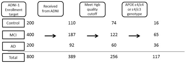
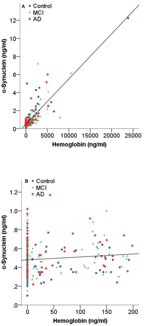
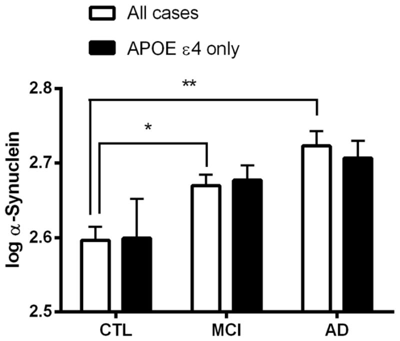
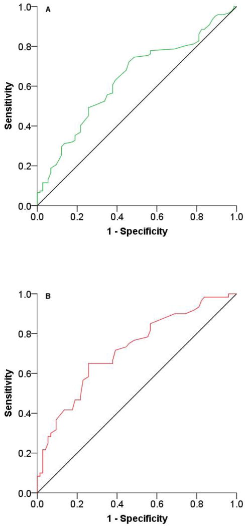
In addition to amyloid-β (Aβ) and tau, α-synuclein, best known for its role in Parkinson's disease (PD), has been suggested to be involved in cognition and pathogenesis of Alzheimer's disease (AD). We investigate the potential of α-synuclein in cerebrospinal fluid (CSF) as a biomarker of cognitive decline in AD, and its prodromal phase, mild cognitive impairment (MCI). Using an established, sensitive Luminex assay, we measured α-synuclein levels in the CSF of a cohort of close to 400 healthy control, MCI, and AD subjects obtained from the Alzheimer's Disease Neuroimaging Initiative (ADNI) and factored in APOE genotype in data analysis. CSF α-synuclein levels were significantly higher in the MCI (p = 0.005) and AD (p < 0.001) groups, compared to controls. However, receiver operating characteristic (ROC) curve analysis suggests that CSF α-synuclein level on its own only offered modest sensitivity (65%) and specificity (74%) as a diagnostic marker of AD, with an area under the curve (AUC) value of 0.719 for AD versus controls. The effect of APOE genotype, if any, was quite subtle. However, there was a significant correlation between α-synuclein and cognition (p = 0.001), with increased α-synuclein levels associated with decreased Mini-Mental State Exam scores. Our results support a role for α-synuclein even in MCI, the early phase of AD, in addition to being a potential contributor in MCI and AD diagnosis or monitoring of disease progression.
Conflict of interest statement
The authors declare no conflict of interest.
Figures

References
-
- Lippa CF, Schmidt ML, Lee VM, Trojanowski JQ. Antibodies to alpha-synuclein detect Lewy bodies in many Down’s syndrome brains with Alzheimer’s disease. Ann Neurol. 1999;45:353–357. - PubMed
-
- Arai Y, Yamazaki M, Mori O, Muramatsu H, Asano G, Katayama Y. Alpha-synuclein-positive structures in cases with sporadic Alzheimer’s disease: morphology and its relationship to tau aggregation. Brain Res. 2001;888:287–296. - PubMed
-
- Olichney JM, Galasko D, Salmon DP, Hofstetter CR, Hansen LA, Katzman R, Thal LJ. Cognitive decline is faster in Lewy body variant than in Alzheimer’s disease. Neurology. 1998;51:351–357. - PubMed
Publication types
MeSH terms
Substances
Grants and funding
- K01 AG030514/AG/NIA NIH HHS/United States
- R01 NS057567/NS/NINDS NIH HHS/United States
- P42 ES004696-5897/ES/NIEHS NIH HHS/United States
- P42 ES004696/ES/NIEHS NIH HHS/United States
- U01 AG024904/AG/NIA NIH HHS/United States
- CAPMC/ CIHR/Canada
- P30 ES007033/ES/NIEHS NIH HHS/United States
- P50 NS062684/NS/NINDS NIH HHS/United States
- R01 AG033398/AG/NIA NIH HHS/United States
- P30 ES007033-6364/ES/NIEHS NIH HHS/United States
- U01 NS082137/NS/NINDS NIH HHS/United States
- P30 AG010129/AG/NIA NIH HHS/United States
- R01 ES019277/ES/NIEHS NIH HHS/United States
- R01 ES016873/ES/NIEHS NIH HHS/United States
LinkOut - more resources
Full Text Sources
Other Literature Sources
Medical
Miscellaneous
