. 2013 Jun 21;113(1):22-31.
doi: 10.1161/CIRCRESAHA.113.301324. Epub 2013 Apr 19.

Apelin-APJ signaling is a critical regulator of endothelial MEF2 activation in cardiovascular development

Affiliations

Apelin-APJ signaling is a critical regulator of endothelial MEF2 activation in cardiovascular development

Yujung Kang et al. Circ Res. .

Abstract

Rationale: The peptide ligand apelin and its receptor APJ constitute a signaling pathway with numerous effects on the cardiovascular system, including cardiovascular development in model organisms such as xenopus and zebrafish.

Objective: This study aimed to characterize the embryonic lethal phenotype of the Apj-/- mice and to define the involved downstream signaling targets.

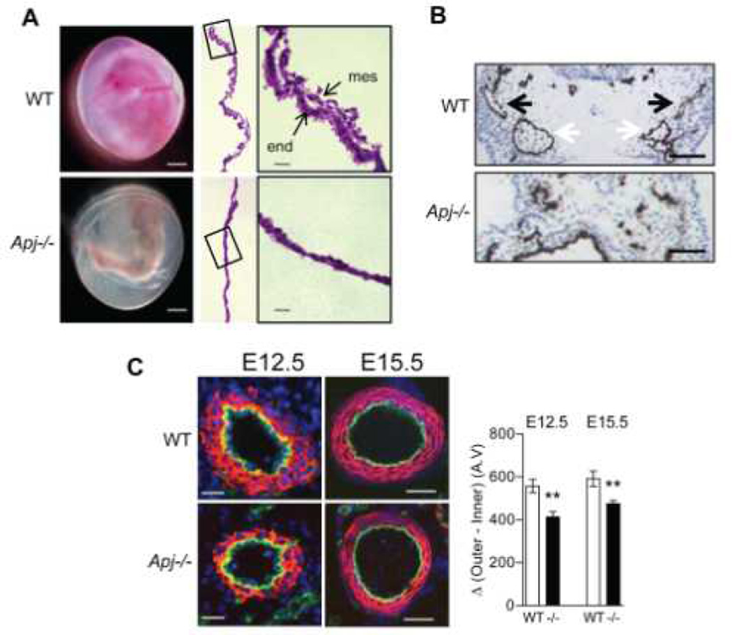
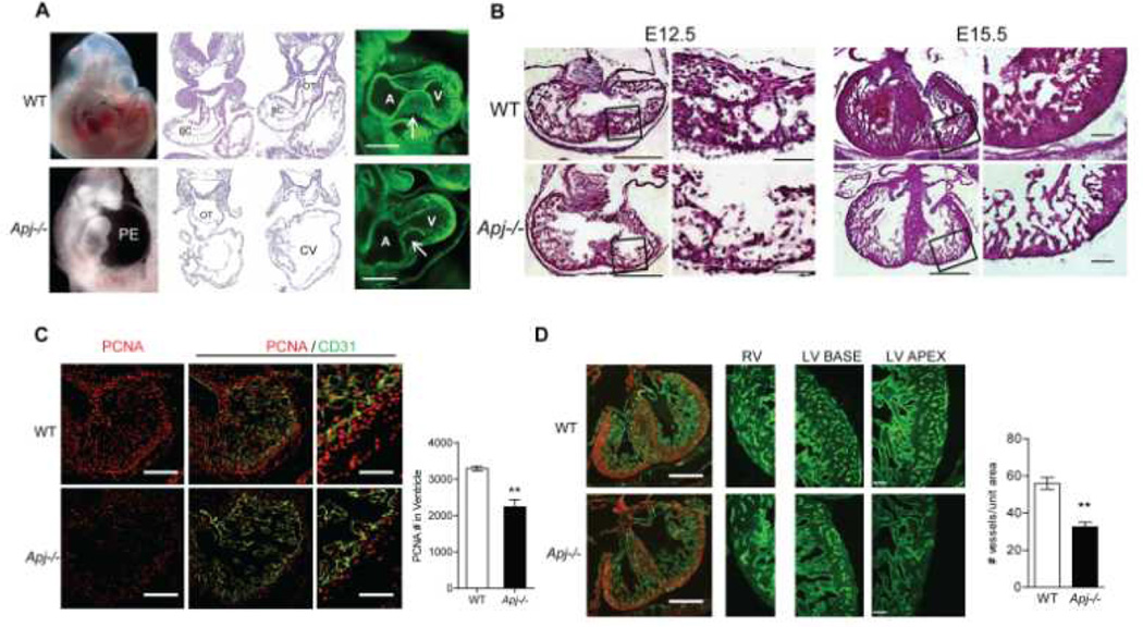
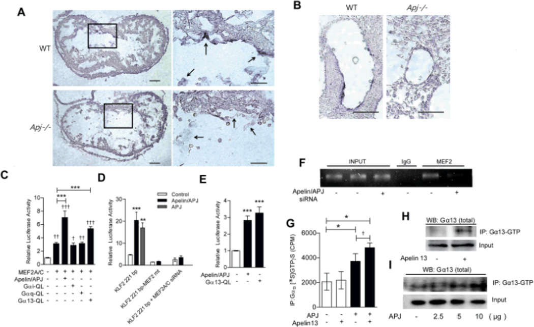
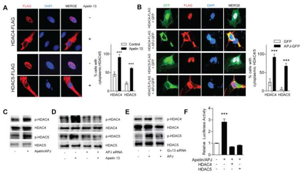
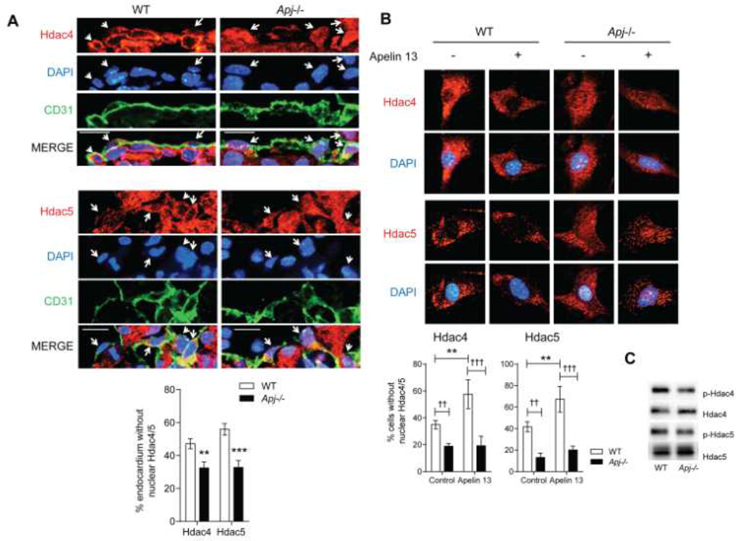
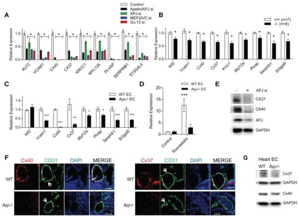
Methods and results: We report the first characterization of the embryonic lethality of the Apj-/- mice. More than half of the expected Apj-/- embryos died in utero because of cardiovascular developmental defects. Those succumbing to early embryonic death had markedly deformed vasculature of the yolk sac and the embryo, as well as poorly looped hearts with aberrantly formed right ventricles and defective atrioventricular cushion formation. Apj-/- embryos surviving to later stages demonstrated incomplete vascular maturation because of a deficiency of vascular smooth muscle cells and impaired myocardial trabeculation and ventricular wall development. The molecular mechanism implicates a novel, noncanonical signaling pathway downstream of apelin-APJ involving Gα13, which induces histone deacetylase (HDAC) 4 and HDAC5 phosphorylation and cytoplasmic translocation, resulting in activation of myocyte enhancer factor 2. Apj-/- mice have greater endocardial Hdac4 and Hdac5 nuclear localization and reduced expression of the myocyte enhancer factor 2 (MEF2) transcriptional target Krüppel-like factor 2. We identify a number of commonly shared transcriptional targets among apelin-APJ, Gα13, and MEF2 in endothelial cells, which are significantly decreased in the Apj-/- embryos and endothelial cells.

Conclusions: Our results demonstrate a novel role for apelin-APJ signaling as a potent regulator of endothelial MEF2 function in the developing cardiovascular system.

Keywords: APJ; Apelin; G proteins; Gα13; HDAC4; HDAC5; MEF2A; MEF2C; developmental biology.


Figures

Figure 1
Figure 1. Vascular defects in Apj−/− embryos
(A) Yolk sac from E10.5 Apj−/− embryos show abnormal yolk sac vasculature compared with wildtype littermates. H&E stain of the yolk sac section shows immature vascular plexus formation. Mes, mesoderm and end, endoderm. White bars indicate 1 mm, black bars indicate 25 µm. (B) CD31 immunohistochemistry of E10.5 Apj−/− embryo section shows defective major vessel development in a subset of Apj−/− embryos. Dorsal aorta (white arrows) and anterior cardinal veins (black arrows) are identified in the wildtype embryo. Bars indicate 100 µm. (C) CD31 and smooth muscle actin (SMA) stain of E12.5 and E15.5 sections show thinner vascular smooth muscle layers surrounding the developing aorta in Apj−/− embryos. SMA staining is shown in red, CD31 staining is shown in green, and DAPI is shown in blue. The wall thickness was calculated from the inner and outer media (SMA positive) circumference in 3 sections from each embryo (n=4–6 embryos per group). Bars indicate 20 µm (E12.5) and 40 µm (E15.5). **P<0.01 vs. wildtype.
Figure 2
Figure 2. Myocardial defects in Apj−/− embryos
(A) E10.5 Apj−/− embryos with cardiac developmental defects, including abnormal chamber development and looping, and large pericardial effusions (PE). H&E stained serial sections show E10.5 Apj−/− embryo with outflow tract (OT) arising from the common ventricle (CV) lacking bulbus cordis (BC), and thin ventricular wall with minimal trabeculation. Right panels show whole mount CD31 staining (green) of E10.5 embryos showing defective atrioventricular cushion formation in Apj−/− embryos (n=6 per group). A, atrium and V, ventricle. Scale bars represent 170 µm. (B) E12.5 and E15.5 Apj−/− embryos have significant thinning of ventricular walls. High magnifications of left ventricular walls are shown in the right panels (n=4–7 per group at each timepoint). Scale bars show 500 µm (low magnification) and 100 µm (high magnification). (C) E12.5 Apj−/− hearts have significant reduction in number of PCNA+ (red) cells (n=4–5 per group). CD31 staining is shown in green. Higher magnifications of left ventricle wall are shown in the right panels. **P<0.01 vs. wildtype. Bars indicate 200 µm (low magnification) and 100 µm (high magnification). D) E15.5 Apj−/− hearts have significantly decreased capillary densities in their ventricular walls compared to their wildtype littermates (n=4–6 per group). CD31 staining is shown in green, SMA staining is shown in red. **P<0.01 vs. wildtype. Scale bars show 500 µm (low magnification) and 100 µm (high magnification).
Figure 3
Figure 3. Apelin and APJ induce MEF2A/C transcriptional activity
(A) In situ hybridization for Klf2 in E10.5 embryos show decreased endocardial (black arrows) Klf2 expression in Apj−/− embryos vs. wildtype littermates (n=3 embryos per group). Scale bars show 50 µm (low magnification) and 100 µm (high magnification). (B) Decreased Klf2 expression in the endothelial layer of the E10.5 dorsal aorta of Apj−/− embryos vs. wildtype littermates. (n=3 embryos per group). Bars indicate 100 µm. (C) MEF2 luciferase reporter containing three tandem MEF2 binding sites is induced by MEF2A/C overexpression in COS7 cells, and induced further by apelin-APJ or constitutively active Gα13 (Gα13-QL), but not by constitutively active Gαi (Gαi-QL) or Gαq (Gαq-QL). ***P<0.001 vs. MEF2A/C alone, P<0.05, ††P<0.01 and †††P<0.001 vs. control. (D) Transfection of apelin and APJ or APJ alone in HUVECs can induce the 221 bp KLF2 promoter driven luciferase reporter containing the MEF2 binding site. This effect is abrogated by either mutation of the MEF2 binding site (MEF2 mt), or by concurrent knockdown of MEF2A and MEF2C (MEF2A/C siRNA). **P<0.01 and ***P<0.001 vs. vector control. (E) Apelin-APJ or Gα13-QL overexpression in HUVECs lead to a significant induction of the 41 basepair KLF2 promoter containing the minimal MEF2 binding site. ***P<0.001 vs. control. (F) ChIP assay shows that MEF2 binding to the KLF2 promoter is reduced in the context of apelin-APJ knockdown in HUVECs. (G) APJ overexpression leads to a significant increase of Gα13 activation in COS7 cells transfected with wildtype Gα13. Stimulation with apelin 13 leads to further activation of Gα13 in APJ transfected cells. *P<0.05 vs. vector control, and P<0.05 vs. APJ without apelin 13 stimulation. (H) Stimulation with apelin 13 leads to an increase in active Gα13 in HUVECs. (I) Transfection of APJ in HUVECs lead to increased levels of active Gα13 in a dosage dependent manner.
Figure 4
Figure 4. Apelin-APJ induces HDAC4/5 phosphorylation and cytoplasmic translocation
(A) Apelin 13 stimulation (1 µM for 1 h) leads to HDAC4 and HDAC5 (red) cytoplasmic translocation in HUVECs transfected with FLAG-tagged HDAC4/5. The percent of cells with cytoplasmic HDAC4/5 in response to apelin 13 stimulation is shown. DAPI (blue) nuclear stain is also shown. ***P<0.001. (B) Overexpression of APJ in HUVECs leads to HDAC4 and HDAC5 cytoplasmic translocation. Cells were transfected with either GFP or APJ-GFP, and co-transfected with FLAG tagged HDAC4 or HDAC5. GFP is shown in green, FLAG staining is shown in red, DAPI nuclear staining is shown in blue. The percent of cells with cytoplasmic HDAC4/5 is shown. ***P<0.001. (C) Overexpression of apelin and APJ in HUVECs lead to increase in HDAC4 and HDAC5 phosphorylation. (D) Stimulation of HUVECs with apelin 13 leads to increased HDAC4 and HDAC5 phosphorylation, which is abrogated in the context of APJ knockdown. (E) Overexpression of APJ in HUVECs leads to increased phosphorylation of HDAC4 and HDAC5, which is decreased with concurrent Gα13 knockdown. (F) Concurrent overexpression of HDAC4 or HDAC5 with apelin-APJ lead to abrogation of apelin-APJ induced activation of the KLF2–41bp luciferase reporter in HUVECs. ***P<0.001 vs. all other conditions.
Figure 5
Figure 5. Apj−/− embryos have significantly higher level of nuclear Hdac4 and Hdac5
(A) Significantly greater number of endocardial cells from E10.5 Apj−/− embryos have nuclear Hdac4 and Hdac5 (red) staining compared to wildtype embryos. DAPI (blue) and CD31 (green) staining are shown (n=4–5 embryos per group). **P<0.01 and ***P<0.001. (B) Isolated heart ECs demonstrate greater percentage of cells lacking nuclear Hdac4 or Hdac5 staining from wildtype mice vs. Apj−/− mice. Only isolated ECs from wildtype mice respond to apelin by translocation of Hdac4 and Hdac5 to the cytoplasm. ECs from Apj−/− demonstrate no response to apelin 13. **P<0.01 vs. no apelin 13 stimulation, ††P<0.01 vs. unstimulated wildtype, †††P<0.001 vs. apelin 13 stimulated wildtype. (C) The level of phosphorylated Hdac4 and Hdac5 are decreased in heart ECs from Apj−/− mice.
Figure 6
Figure 6. Shared targets of apelin-APJ, Gα13, and MEF2 in ECs are downregulated in Apj−/− embryos and isolated ECs
(A) Knockdown of apelin/APJ, APJ, MEF2A/C or Gα13 in HUVECs leads to decreased mRNA expression of validated and putative MEF2 target genes. *P<0.05 for each knockdown condition compared to control. (B) Transcripts decreased in HUVECs subjected to apelin-APJ, APJ, MEF2A/C or Gα13 knockdown are also significantly decreased in E10.5 Apj−/− embryos. *P<0.05 vs. wildtype. (C) Isolated heart ECs from Apj−/− mice demonstrate decreased expression of gene transcripts identified from the microarray analyses. *P<0.05, **P<0.01 and ***P<0.001. (D) KLF2 expression is significantly induced by rosuvastatin in isolated heart ECs from wildtype mice but not from Apj−/− mice. ***P<0.001 vs. all other conditions. (E) Expression of CX37 and CX40 are decreased in HUVECs subjected to APJ knockdown. (F) Cx37 and Cx40 in CD31 staining ECs of the dorsal aorta (white arrows) are decreased in E10.5 Apj−/− embryos (n=4–5 embryos per group). Cx37/40 staining is shown in red, CD31 is shown in green, and DAPI is shown in blue. Bar indicates 80 µm. (G) Decreased expression of Cx37 and Cx40 in heart ECs of Apj−/− mice.

Comment in

References

    1. Scimia MC, Hurtado C, Ray S, Metzler S, Wei K, Wang J, Woods CE, Purcell NH, Catalucci D, Akasaka T, Bueno OF, Vlasuk GP, Kaliman P, Bodmer R, Smith LH, Ashley E, Mercola M, Brown JH, Ruiz-Lozano P. Apj acts as a dual receptor in cardiac hypertrophy. Nature. 2012;488:394–398. - PMC - PubMed
    1. Chun HJ, Ali ZA, Kojima Y, Kundu RK, Sheikh AY, Agrawal R, Zheng L, Leeper NJ, Pearl NE, Patterson AJ, Anderson JP, Tsao PS, Lenardo MJ, Ashley EA, Quertermous T. Apelin signaling antagonizes ang ii effects in mouse models of atherosclerosis. The Journal of clinical investigation. 2008;118:3343–3354. - PMC - PubMed
    1. Kim J, Kang Y, Kojima Y, Lighthouse JK, Hu X, Aldred MA, McLean DL, Park H, Comhair SA, Greif DM, Erzurum SC, Chun HJ. An endothelial apelin-fgf link mediated by mir-424 and mir-503 is disrupted in pulmonary arterial hypertension. Nature medicine. 2013;19:74–82. - PMC - PubMed
    1. Chandra SM, Razavi H, Kim J, Agrawal R, Kundu RK, de Jesus Perez V, Zamanian RT, Quertermous T, Chun HJ. Disruption of the apelin-apj system worsens hypoxia-induced pulmonary hypertension. Arteriosclerosis, thrombosis, and vascular biology. 2011;31:814–820. - PMC - PubMed
    1. O'Dowd BF, Heiber M, Chan A, Heng HH, Tsui LC, Kennedy JL, Shi X, Petronis A, George SR, Nguyen T. A human gene that shows identity with the gene encoding the angiotensin receptor is located on chromosome 11. Gene. 1993;136:355–360. - PubMed

Publication types

MeSH terms