. 2013 Jun;12(6):576-83.
doi: 10.1038/nmat3627. Epub 2013 Apr 21.

Nano-analytical electron microscopy reveals fundamental insights into human cardiovascular tissue calcification

Affiliations

Nano-analytical electron microscopy reveals fundamental insights into human cardiovascular tissue calcification

Sergio Bertazzo et al. Nat Mater. 2013 Jun.

Abstract

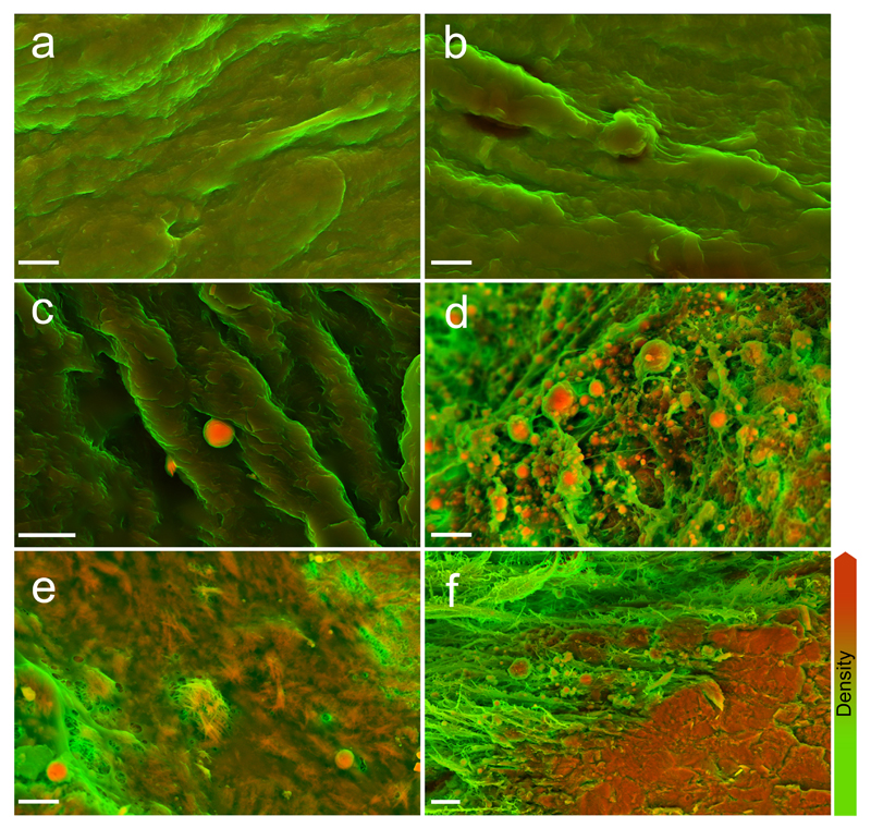
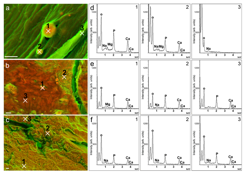
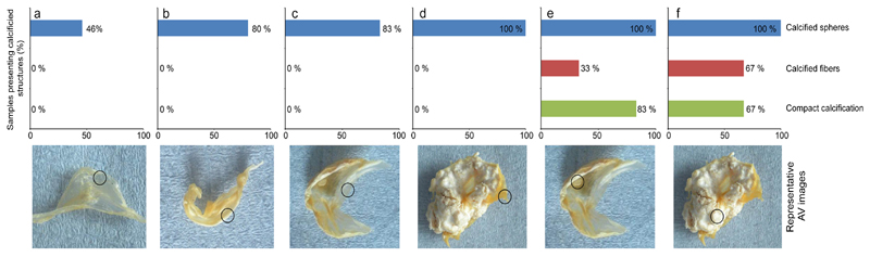
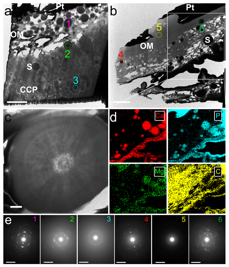
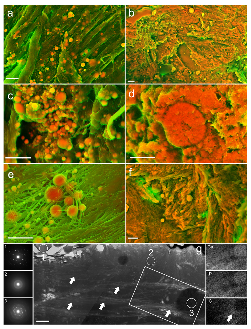
The accumulation of calcified material in cardiovascular tissue is thought to involve cytochemical, extracellular matrix and systemic signals; however, its precise composition and nanoscale architecture remain largely unexplored. Using nano-analytical electron microscopy techniques, we examined valves, aortae and coronary arteries from patients with and without calcific cardiovascular disease and detected spherical calcium phosphate particles, regardless of the presence of calcific lesions. We also examined lesions after sectioning with a focused ion beam and found that the spherical particles are composed of highly crystalline hydroxyapatite that crystallographically and structurally differs from bone mineral. Taken together, these data suggest that mineralized spherical particles may play a fundamental role in calcific lesion formation. Their ubiquitous presence in varied cardiovascular tissues and from patients with a spectrum of diseases further suggests that lesion formation may follow a common process. Indeed, applying materials science techniques to ectopic and orthotopic calcification has great potential to lend critical insights into pathophysiological processes underlying calcific cardiovascular disease.


Conflict of interest statement

Financial Disclosure

The authors declare no competing financial interests.

Figures

Figure 1
Figure 1. Density-dependent color scanning electron micrographs of human aortic valve (AV) tissue.
Micrographs were coloured in post-processing by combing images acquired by secondary and backscatter electron detectors (details and original images are in Supplementary Fig. 3). The orange colour identifies denser material, while structures that appear green are less dense. a, b Surface of AV rejected for transplant because of physical damage. Samples lacked macroscopic calcific lesions and were judged not to be atheromic. c Surface of AV free from macroscopically observable calcific lesions, but presenting dense spherical particles. d Calcific lesion on AV with notable dense spherical particles. e Calcific lesion on AV, presenting dense spherical particles and fibers. f Calcific lesion on AV, presenting dense spherical particles and compact material. Scale bar = 3 µm.
Figure 2
Figure 2. Elemental analyses of dense structures identified on aortic valves (AV).
Density-dependent colour scanning electron micrographs of AV presenting (a) dense spherical particles, (b) dense fibers and (c) compact material. d, e and f depict corresponding energy-dispersive X-ray spectra collected at the numbered sites indicated on micrographs a, b and c, respectively. Scale bar: 2 = µm.
Figure 3
Figure 3. Histogram describing the prevalence of dense calcium- and phosphorus-containing structures on the surface of aortic valves (AV).
Representative images of AV are categorized by presence and extensiveness of calcific lesions. Sampled regions indicated in circles. Category a is comprised of AV that did not present any macroscopically observable calcific lesions on AV or in surrounding tissue (aorta and coronary arteries). b represents AV without macroscopically observable calcification on AV, but with macroscopically observable calcific lesions on surrounding tissues. c includes analyses carried out on non-calcified areas of AV which presented calcific lesions in other areas of the AV. d includes analyses carried out on non-calcified areas of AV which were heavily calcified in other areas. e identifies structures on macroscopically observable calcific lesions. f includes analyses carried out on calcific lesions from heavily calcified AV. Approximately 350 images were obtained from each of the 55 regions examined in AV from 32 patients.
Figure 4
Figure 4. Transmission electron microscopy (TEM) images, energy-dispersive X-ray spectroscopy (EDS) mapping, and selected area electron diffraction (SAED) analyses of aortic valve (AV) tissues sectioned with a focussed ion beam (FIB).
a/b TEM images show spherical particles (S) trapped in compact calcium phosphate (CCP) and organic matrices (OM). AV surfaces are coated in a dense layer of platinum (Pt) utilized in sample preparation for FIB. Scale bar = 2 µm. c High-resolution TEM of calcium- and phosphorus-containing spherical particle on the surface of a calcific lesion. Scale bar = 0.2 µm. d Elemental mapping of calcium (Ca), phosphorus (P), magnesium (Mg) and carbon (C) in the region inside the highlighted area in b (original images and elemental maps of gallium, the ion source utilized by the FIB, and platinum are included in Supplementary Figs. 5/6). e SAED obtained from each of the sections inside the numbered circles in a and b. Scale bar = 0.05 nm-1. Note that spherical particles produce diffraction patterns typical of highly crystalline material, while the surrounding compact material yielded diffraction patterns typical of a poorly crystalline material.
Figure 5
Figure 5. Density-dependent colour scanning electron micrographs of mitral valve, aorta and coronary artery, and transmission electron microscopy (TEM), selected area electron diffraction (SAED) and energy dispersion X-ray spectroscopy (EDS) mapping of calcific lesions on coronary artery tissue sectioned using a focussed ion beam (FIB).
a Mitral valve tissue with notable dense spheres. b Calcific lesion on mitral valve tissue presenting dense spherical particles and compact material. c Aortic tissue with notable dense spherical particles. d Calcific lesion on aortic tissue presenting dense spherical particles and compact material. e Coronary artery tissue with dense spherical particles. f Calcific lesion on coronary artery presenting dense spherical particles and compact material. Further examples of dense structures and dense fibers are provided in Supplementary Fig. 7. Scale bar = 3 µm. g TEM of a calcific lesion on coronary artery tissue shown with SAED images of the numbered regions (left-hand column) and elemental mapping by EDS (right-hand column) of areas highlighted in the central image. Arrows point to collagen fibers, which are notably not intimately associated with the mineralized spherical particles. Scale bar = 2 µm.

Comment in

References

    1. Butler D. UN targets top killers. Nature. 2011:260–261. - PubMed
    1. Rajamannan NM, et al. Human aortic valve calcification is associated with an osteoblast phenotype. Circulation. 2003;107:2181–2184. - PMC - PubMed
    1. Lindroos M, Kupari M, Heikkila J, Tilvis R. Prevalence of aortic valve abnormalities in the elderly: an echocardiographic study of a random population sample. J Am Coll Cardiol. 1993;21:1220–1225. - PubMed
    1. Nkomo VT, et al. Burden of valvular heart diseases: a population-based study. Lancet. 2006;368:1005–1011. - PubMed
    1. Roger VL, et al. Heart Disease and Stroke Statistics—2011 Update. Circulation. 2011;123:e18–e209. - PMC - PubMed

Publication types