. 2013 Jun 21;288(25):18172-83.
doi: 10.1074/jbc.M112.407296. Epub 2013 Apr 19.

Endoplasmic reticulum stress regulates rat mandibular cartilage thinning under compressive mechanical stress

Affiliations

Endoplasmic reticulum stress regulates rat mandibular cartilage thinning under compressive mechanical stress

Huang Li et al. J Biol Chem. .

Abstract

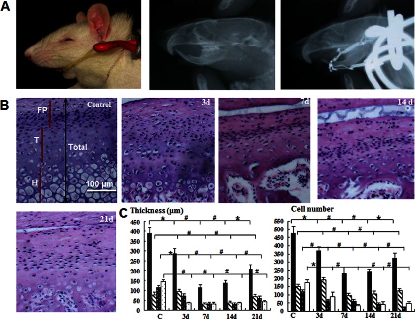
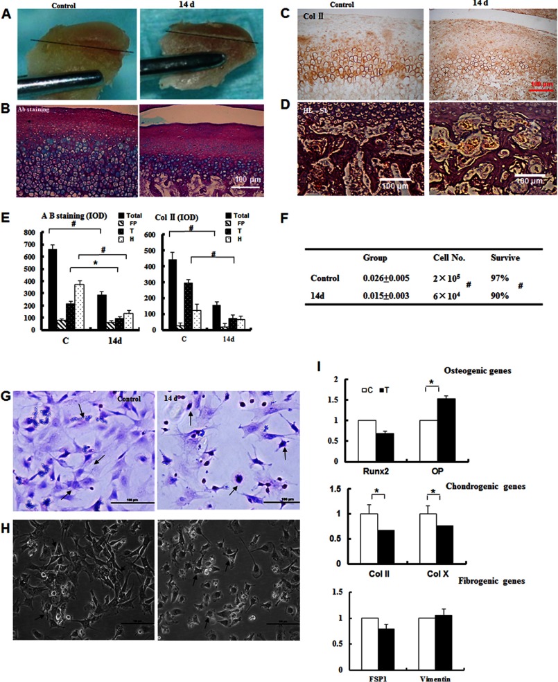
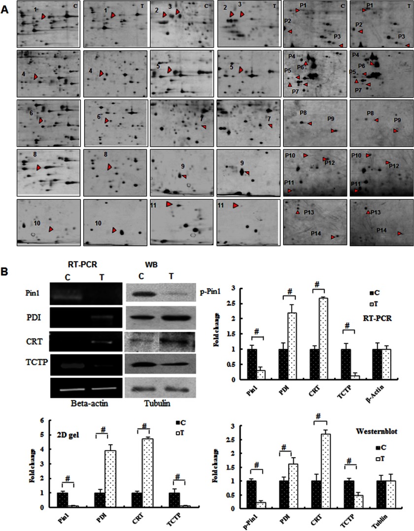
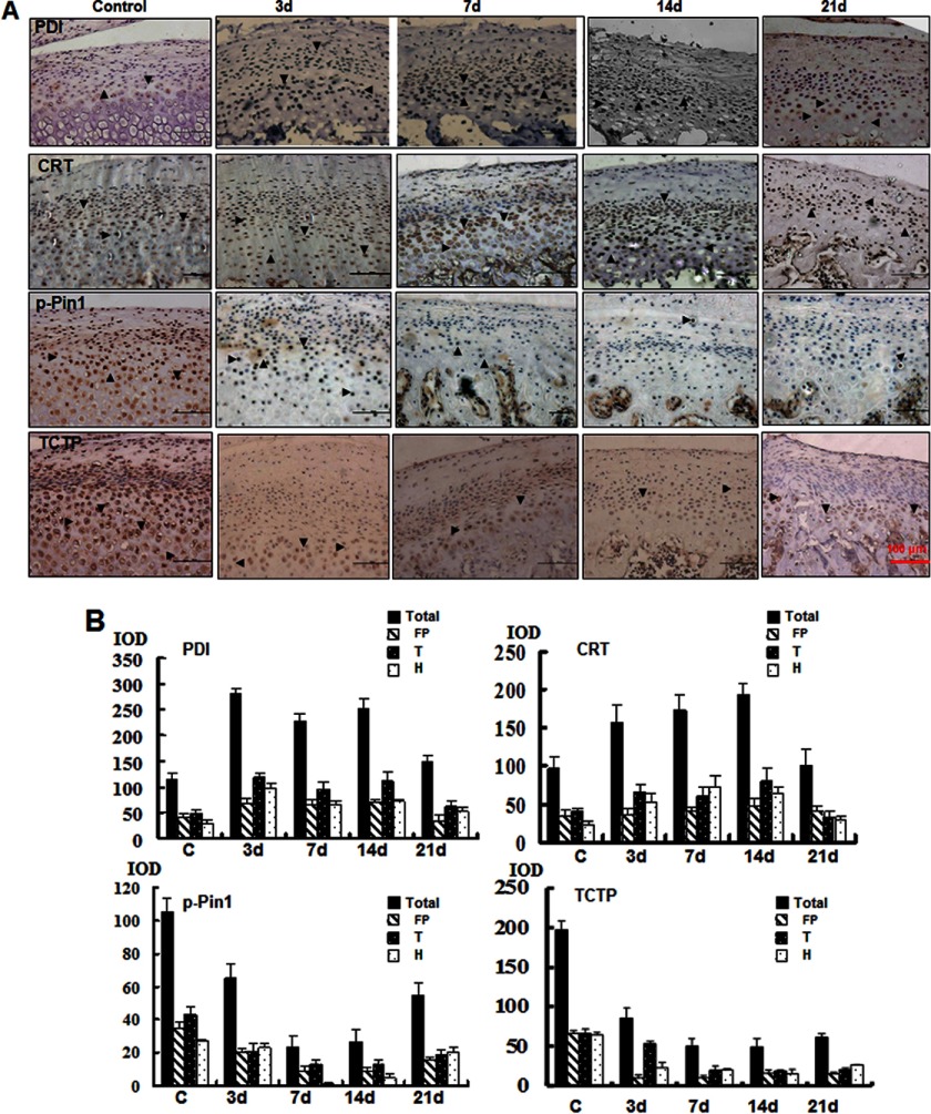
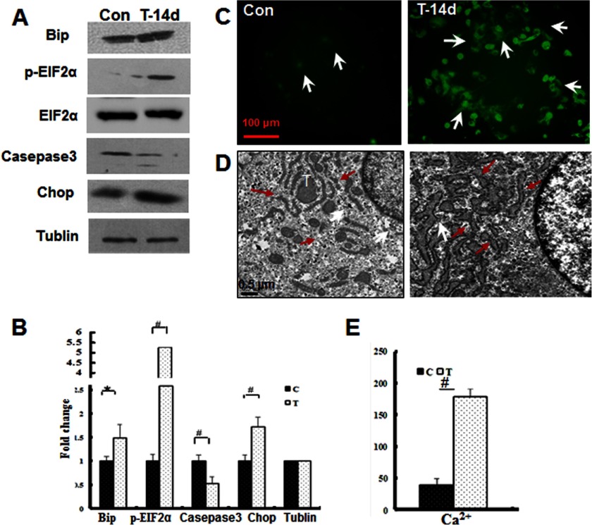
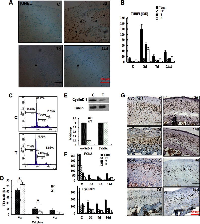
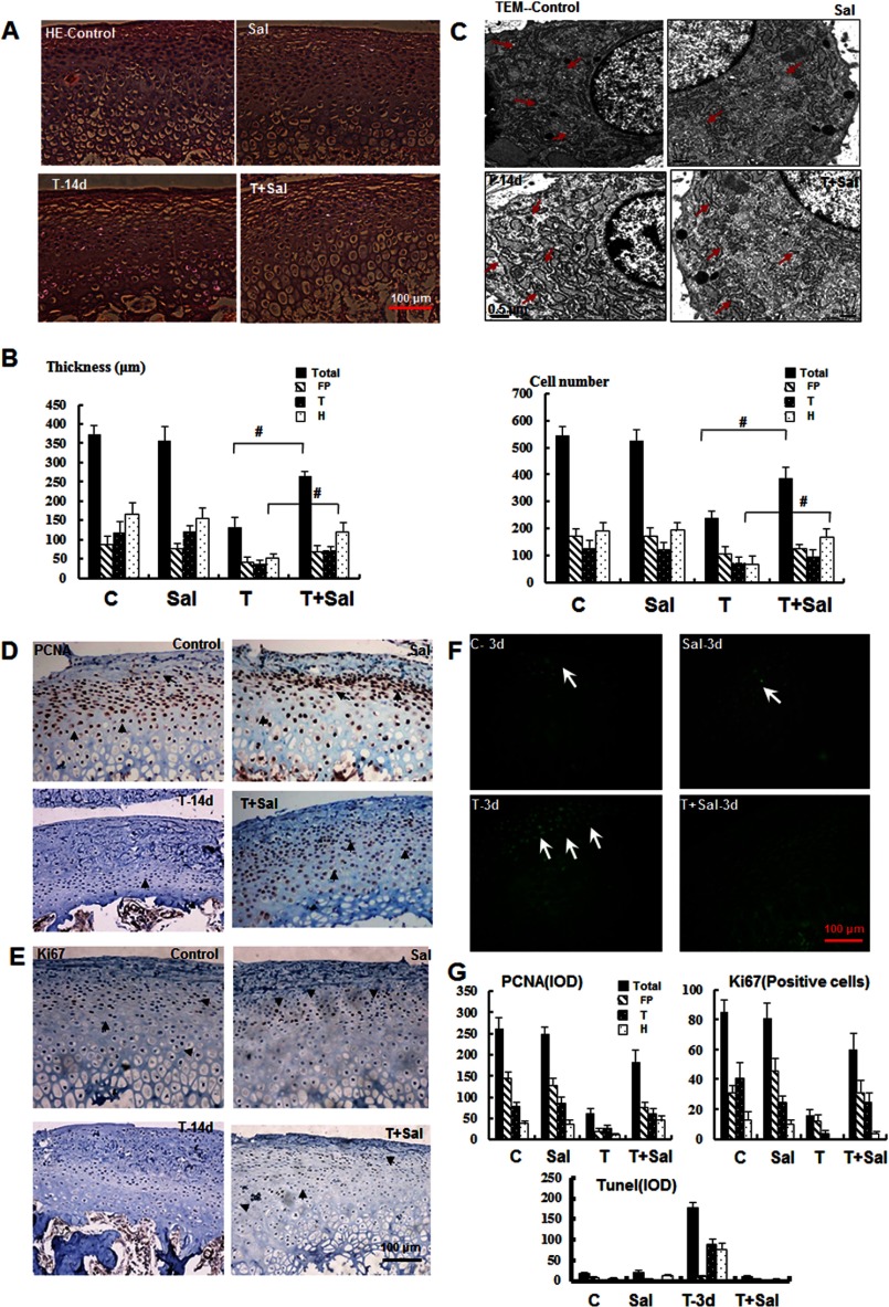
Compressive mechanical stress-induced cartilage thinning has been characterized as a key step in the progression of temporomandibular joint diseases, such as osteoarthritis. However, the regulatory mechanisms underlying this loss have not been thoroughly studied. Here, we used an established animal model for loading compressive mechanical stress to induce cartilage thinning in vivo. The mechanically stressed mandibular chondrocytes were then isolated to screen potential candidates using a proteomics approach. A total of 28 proteins were identified that were directly or indirectly associated with endoplasmic reticulum stress, including protein disulfide-isomerase, calreticulin, translationally controlled tumor protein, and peptidyl-prolyl cis/trans-isomerase protein. The altered expression of these candidates was validated at both the mRNA and protein levels. The induction of endoplasmic reticulum stress by mechanical stress loading was confirmed by the activation of endoplasmic reticulum stress markers, the elevation of the cytoplasmic Ca(2+) level, and the expansion of endoplasmic reticulum membranes. More importantly, the use of a selective inhibitor to block endoplasmic reticulum stress in vivo reduced the apoptosis observed at the early stages of mechanical stress loading and inhibited the proliferation observed at the later stages of mechanical stress loading. Accordingly, the use of the inhibitor significantly restored cartilage thinning. Taken together, these results demonstrated that endoplasmic reticulum stress is significantly activated in mechanical stress-induced mandibular cartilage thinning and, more importantly, that endoplasmic reticulum stress inhibition alleviates this loss, suggesting a novel pharmaceutical strategy for the treatment of mechanical stress-induced temporomandibular joint diseases.

Keywords: Cartilage; Chondrocytes; ER Stress; Mechanotransduction; Proteomics.


Figures

FIGURE 1.
FIGURE 1.
Compressive mechanical stress induces mandibular cartilage thinning. A, an appliance that loaded 40 g of force on each side was used to move the mandible posteriorly and superiorly. B, H&E staining (magnification ×100) of samples subjected or not subjected to mechanical stress for the number of days indicated. P, proliferative zone; T, transition zone; H, hypertrophic zone. C, quantitative analysis of cartilage thickness and the cell number from the samples shown in B (*, p < 0.05; #, p < 0.01). Error bars, S.D.
FIGURE 2.
FIGURE 2.
Isolation and characterization of mechanically stressed chondrocytes. A, chondrocytes were isolated from the covered cartilage (above black lines) in control and 14-day samples. B, the cartilage was untreated or treated for 14 days, and the samples were subsequently stained with Alcian blue (Ab) (magnification ×100). C, the samples from C were subjected to collagen II (Col II) immunostaining. D, expansion of the bone marrow space and subchondral bone erosion were observed (magnification ×100). E, the quantitative analysis from B and C confirmed the observations in the extracellular matrix and the loss of collagen II immunostaining. *, p < 0.05; #, p < 0.01. F, the cell numbers and the survival rate were reduced in the 14-day group (#, p < 0.01). G, the primary chondrocyte cultures were evaluated by H&E staining (magnification ×100). H, the primary chondrocytes cultures were examined by inverted phase-contrast microscopy (magnification ×100). I, real-time quantitative PCR analysis of osteogenic, chondrogenic, and fibrogenic genes as indicated. Error bars, S.D.
FIGURE 3.
FIGURE 3.
Identification of differentially expressed proteins by proteomic analysis. A, the protein spots are indicated with arrows and numbers corresponding to the proteins listed in supplemental Table 1. B, RT-PCR and Western blot (WB) analyses of the proteins, as indicated, and quantification of the -fold changes observed in the two-dimensional gel, RT-PCR, and Western blot analyses of these proteins (#, p < 0.01). Error bars, S.D.
FIGURE 4.
FIGURE 4.
Immunohistochemical analysis of time-dependent changes in the differential expression of proteins in vivo. A, immunohistochemical analysis of the proteins from samples treated for different numbers of days as indicated (magnification ×100). B, quantification of the changes in the expression of these proteins. The integral optical density (IOD) was analyzed by Image-Pro Plus. Error bars, S.D.
FIGURE 5.
FIGURE 5.
Induction of ERS in response to mechanical stress. A, Western blot analysis of the ERS marker proteins Bip, phospho-eIF2α, cleaved caspase-3, and CHOP from the control (Con) or mechanical stress-treated (T-14d) samples. B, quantification of the -fold changes in the proteins shown in A (#, p < 0.01; *, p < 0.05). C, effect of mechanical stress on free Ca2+ levels in mandibular chondrocytes as assessed by immunofluorescent staining. Free Ca2+ flux was visualized with a specific indicator (white arrowhead) (magnification ×100). D, TEM (magnification ×2000) revealed that the ER was expanded under mechanical stress (red arrows). E, the quantification of the changes in the Ca2+ levels is shown (#, p < 0.01). Error bars, S.D.
FIGURE 6.
FIGURE 6.
Mechanical stress-induced apoptosis, cell cycle arrest, and proliferation inhibition at different loading times. A, apoptosis was examined by TUNEL staining. B, quantification of A by integral optical density (IOD). C, cell cycle analysis of mandibular chondrocytes treated (T) or untreated (C) with mechanical stress for 14 days. D, the DNA histogram revealed a marked reduction in the S-phase percentage and increase in the G1-phase percentage (*, p < 0.05). E, Western blot analysis of cyclin D1 expression and quantification of the changes in the samples treated (T) or untreated (C) with mechanical stress for 14 days. F and G, immunohistochemical analysis of PCNA and cyclin D1 in samples treated for the indicated number of days. Error bars, S.D.
FIGURE 7.
FIGURE 7.
Inhibition of ERS alleviates mandibular cartilage thinning. A, H&E staining of the mandibular cartilage from samples treated as indicated for 14 days (magnification ×100). Sal, salubrinal. B, quantification of the results shown in A for the cartilage thickness and cell number in different zones as indicated (#, p < 0.01). C, TEM (magnification ×2000) revealed that the expanded ER membrane under mechanical stress was improved after inhibition of ERS (red arrowhead). D and E, immunohistochemical analysis of PCNA and Ki67 in samples treated for 14 days. F, TUNEL staining of samples treated as indicated for 3 days. G, quantification of the observed proliferation and apoptosis. Error bars, S.D.

References

    1. Sicher H. (1947) The growth of the mandible. Am. J. Orthod. 43, 123–128 - PubMed
    1. Kiliaridis S., Thilander B., Kjellberg H., Topouzelis N., Zafiriadis A. (1999) Effect of low masticatory function on condylar growth. A morphometric study in the rat. Am. J. Orthod. Dentofacial Orthop. 116, 121–125 - PubMed
    1. Murray R. C., Zhu C. F., Goodship A. E., Lakhani K. H., Agrawal C. M., Athanasiou K. A. (1999) Exercise affects the mechanical properties and histological appearance of equine articular cartilage. J. Orthop. Res. 17, 725–731 - PubMed
    1. Kuroda S., Tanimoto K., Izawa T., Fujihara S., Koolstra J. H., Tanaka E. (2009) Biomechanical and biochemical characteristics of the mandibular condylar cartilage. Osteoarthr. Cartil. 17, 1408–1415 - PubMed
    1. Wadhwa S., Kapila S. (2008) TMJ disorders. Future innovations in diagnostics and therapeutics. J. Dent. Educ. 72, 930–947 - PMC - PubMed

Publication types