. 2013 May 31;288(22):15614-27.
doi: 10.1074/jbc.M112.443507. Epub 2013 Apr 19.

The choline-binding protein PspC of Streptococcus pneumoniae interacts with the C-terminal heparin-binding domain of vitronectin

Affiliations

The choline-binding protein PspC of Streptococcus pneumoniae interacts with the C-terminal heparin-binding domain of vitronectin

Sylvia Voss et al. J Biol Chem. .

Abstract

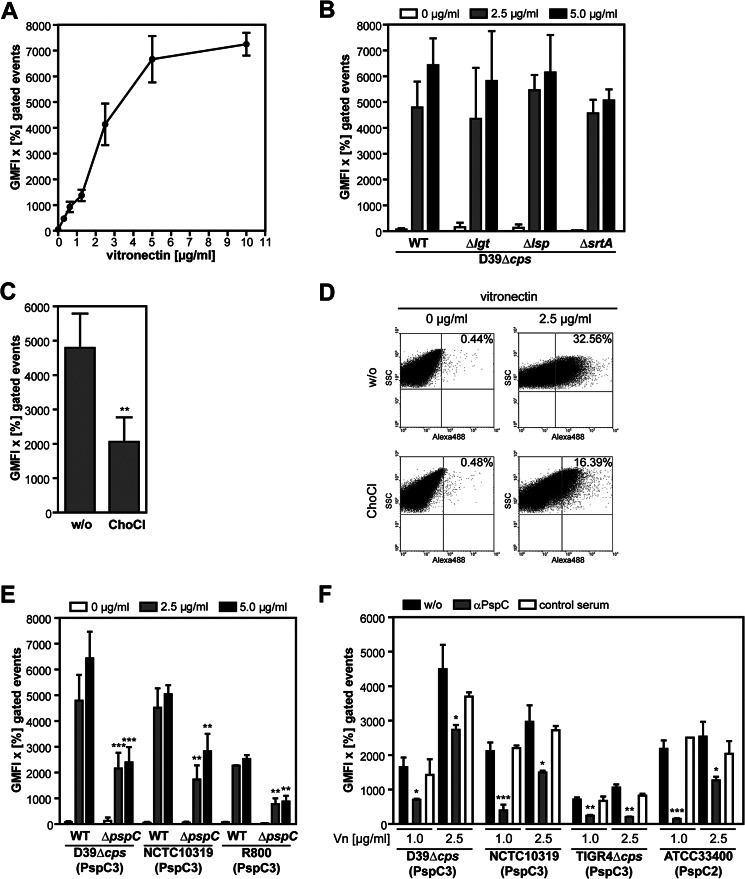
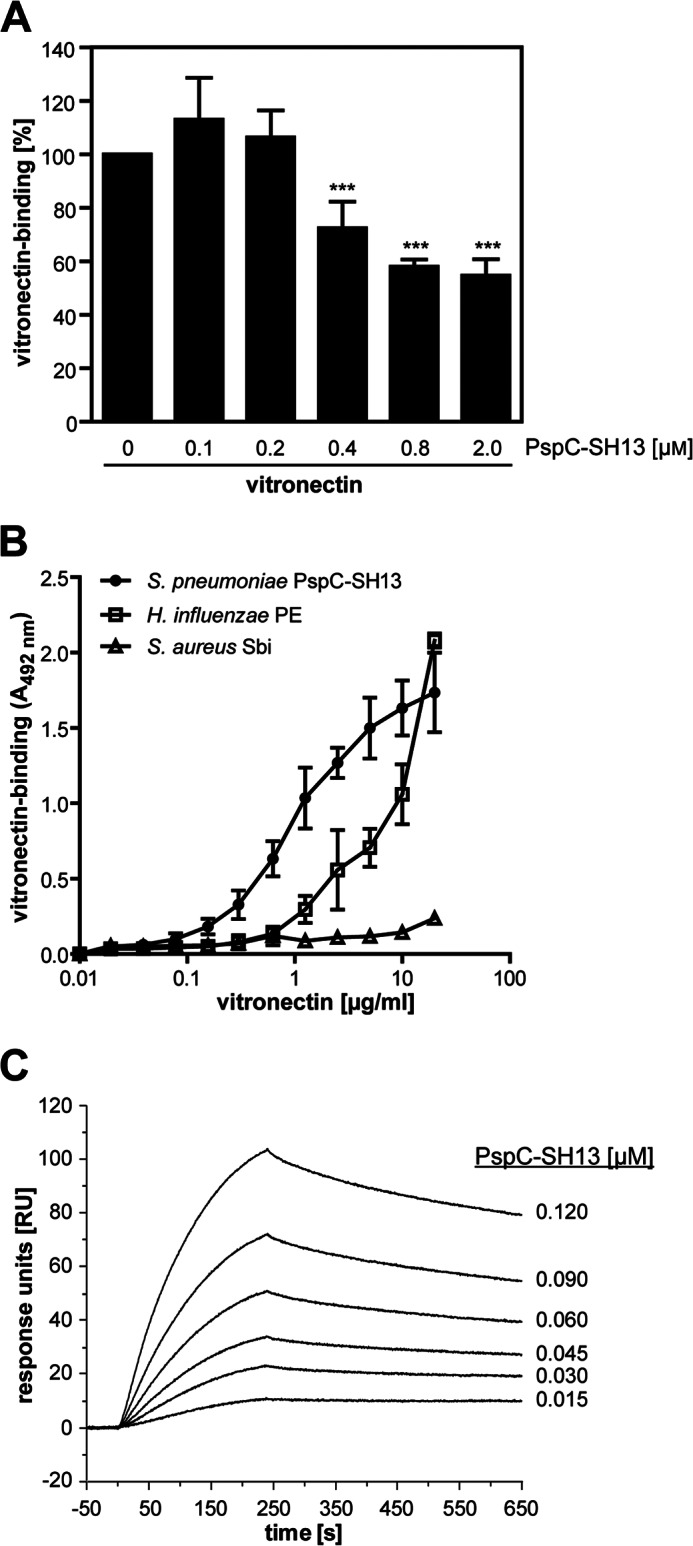
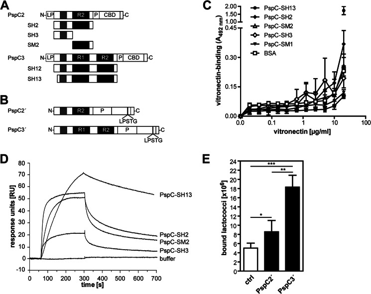
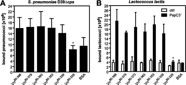
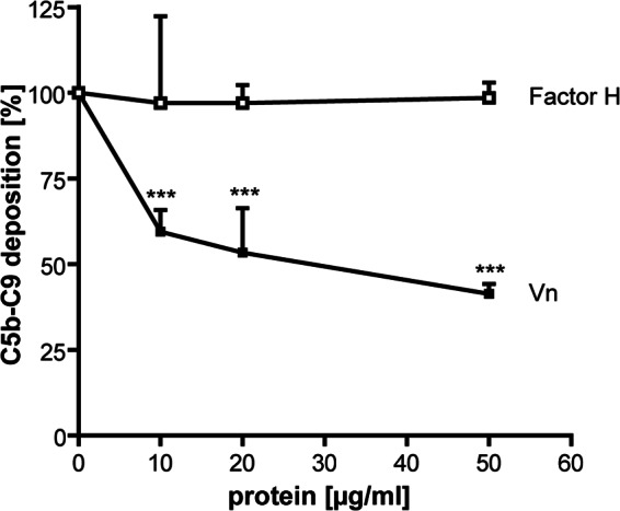
Adherence of Streptococcus pneumoniae is directly mediated by interactions of adhesins with eukaryotic cellular receptors or indirectly by exploiting matrix and serum proteins as molecular bridges. Pneumococci engage vitronectin, the human adhesive glycoprotein and complement inhibitor, to facilitate attachment to epithelial cells of the mucosal cavity, thereby modulating host cell signaling. In this study, we identified PspC as a vitronectin-binding protein interacting with the C-terminal heparin-binding domain of vitronectin. PspC is a multifunctional surface-exposed choline-binding protein displaying various adhesive properties. Vitronectin binding required the R domains in the mature PspC protein, which are also essential for the interaction with the ectodomain of the polymeric immunoglobulin receptor and secretory IgA. Consequently, secretory IgA competitively inhibited binding of vitronectin to purified PspC and to PspC-expressing pneumococci. In contrast, Factor H, which binds to the N-terminal part of mature PspC molecules, did not interfere with the PspC-vitronectin interaction. Using a series of vitronectin peptides, the C-terminal heparin-binding domain was shown to be essential for the interaction of soluble vitronectin with PspC. Binding experiments with immobilized vitronectin suggested a region N-terminal to the identified heparin-binding domain as an additional binding region for PspC, suggesting that soluble, immobilized, as well as cellularly bound vitronectin possesses different conformations. Finally, vitronectin bound to PspC was functionally active and inhibited the deposition of the terminal complement complex. In conclusion, this study identifies and characterizes (on the molecular level) the interaction between the pneumococcal adhesin PspC and the human glycoprotein vitronectin.

Keywords: Adhesin; Bacterial Pathogenesis; Complement; Extracellular Matrix; Pneumococci; Protein-Protein Interactions; PspC; Surface Plasmon Resonance (SPR); Virulence Factors.


Figures

FIGURE 1.
FIGURE 1.
Binding of human vitronectin to the pneumococcal surface is mediated by PspC. Recruitment of soluble vitronectin to untreated or pretreated wild-type pneumococci or isogenic mutants (100 μl of 1 × 10⁸ bacteria) was determined by flow cytometry. Bound vitronectin was detected using anti-human vitronectin antibodies, followed by an AlexaFluor488-conjugated anti-rabbit IgG. The results were expressed as GMFI × percentage of AlexaFluor488-labeled and gated bacteria. The mean values of at least three independent experiments are shown with error bars corresponding to S.D. **, p ≤ 0.01; ***, p ≤ 0.001. Representative dot plots are shown in supplemental Fig. S1. A, dose-dependent binding of soluble vitronectin to S. pneumoniae D39Δcps. B, binding of soluble vitronectin to S. pneumoniae D39Δcps (WT) and its isogenic mutants D39ΔcpsΔlgt, D39ΔcpsΔlsp, or D39ΔcpsΔsrtA. C–D, binding of soluble vitronectin to pneumococci devoid of choline-binding proteins. C, S. pneumoniae D39Δcps were incubated with vitronectin (2.5 μg/ml) in the absence (w/o) or presence of 5% choline chloride (ChoCl). D, vitronectin bound to the pneumococcal surface in the absence (w/o) or presence of choline chloride is shown as dot plots of a representative flow cytometric analysis. The control dot plots show the GMFI × percentage of gated bacteria in the absence of vitronectin but after incubation with the antibodies. E, binding of soluble vitronectin to various pneumococcal strains, represented by low encapsulated (NCTC10319) or nonencapsulated (D39Δcps, R800) pneumococci, and their isogenic ΔpspC mutants. The values for the vitronectin binding to D39Δcps were transferred from B. F, inhibition of vitronectin binding to pneumococci expressing different PspC subtypes with a mouse anti-PspC serum or mouse control serum. Vitronectin was used at a concentration of 1 and 2.5 μg/ml, respectively.
FIGURE 2.
FIGURE 2.
Vitronectin binding to PspC subtype 3. A, competitive inhibition of vitronectin binding to pneumococci using a derivative of PspC subtype 3. S. pneumoniae D39Δcps (100 μl of 1 × 10⁸ bacteria) were incubated with vitronectin (1 μg/ml), and binding was competitively inhibited by the addition of increasing concentrations of PspC-SH13 (0–2 μm). Binding of vitronectin was determined by flow cytometry (for dot plots of a representative flow cytometric analysis, see supplemental Fig. S2), and data show the percentage of binding relative to vitronectin binding to pneumococci in the absence of protein. The mean values of at least three independent experiments and the S.D. are shown. ***, p ≤ 0.001. B, dose-dependent binding of soluble vitronectin to PspC-SH13. PspC-SH13, S. aureus Sbi, or H. influenzae PE were coated on microtiter plates (each 100 μl of 5 μg/ml) and incubated with increasing concentrations of vitronectin (0–25 μg/ml in 100 μl). Bound vitronectin was detected with anti-human vitronectin antibodies, followed by incubation with HRP-conjugated anti-goat antibodies. 1,2-Phenylenediamine dihydrochloride was used as a substrate, and the absorbance was measured at 492 nm. Results are the mean values ± S.D. of three independent experiments. C, surface plasmon resonance measurements of PspC-SH13 binding to immobilized vitronectin. Vitronectin was immobilized on a CM5 biosensor chip to a final rate of 350 response units (RU). PspC-SH13 was used as an analyte at a flow rate of 10 μl/min, and the affinity surface was regenerated between subsequent sample injections with 12.5 mm sodium hydroxide. PspC-SH13 showed a dose-dependent binding to vitronectin expressed in arbitrary response units. A control flow cell was used to subtract nonspecific signals.
FIGURE 3.
FIGURE 3.
Vitronectin preferentially interacts with PspC proteins of subtype 3. A, schematic model of the PspC subtypes 2 and 3 produced as N-terminally His6-tagged protein derivatives in E. coli. B, schematic model of the PspC′-LPSTG fusion proteins for the heterologous expression of PspC on the surface of L. lactis. Factor H-binding domain of PspC is shown in dark gray, and the R domains (R1 and R2) of the different PspC variants are depicted in black. Each R domain contains the hexapeptide-binding motif (Y/R)RNYPT for the SC of the pIgR and sIgA, respectively. LP, Leader peptide; CBD, choline-binding domain; P, proline-rich sequence; R, repeat domain. C, binding of vitronectin to immobilized PspC derivatives. PspC subtype 2 (PspC-SH2) or part structures of the mature PspC protein (PspC-SH3 or -SM2) were immobilized on microtiter plates (each 100 μl of 5 μg/ml) and incubated with increasing concentrations of vitronectin (0–25 μg/ml in 100 μl). PspC subtype 3 (PspC-SH13) is shown as the control, and the value was transferred from Fig. 2. Binding of vitronectin was detected as described in Fig. 2. Results are the mean values ± S.D. of three independent experiments. D, binding of PspC derivatives to immobilized vitronectin measured by surface plasmon resonance. PspC-SH13 (representing subtype 3; 0.09 μm), PspC-SH2 (representing subtype 2; 1 μm), the mature N-terminal domain of PspC (PspC-SH3; 4 μm), and one R domain (PspC-SM2; 4 μm), respectively, were used as analytes at a flow rate of 10 μl/min, and the affinity surface was regenerated between subsequent sample injections of proteins with 12.5 mm sodium hydroxide. The amount of PspC binding to immobilized vitronectin is shown in arbitrary response units (RU). E, binding of PspC-expressing lactococci to immobilized vitronectin. PspC expression in L. lactis was induced, and 2 × 10⁸ recombinant lactococci were labeled with FITC. Binding of FITC-labeled bacteria to immobilized vitronectin (50 μl of 10 μg/ml) was measured at 485/538 nm (excitation/emission), and the number of bound bacteria was calculated. The mean values ± S.D. of at least three independent experiments performed in triplicates are shown. *, p ≤ 0.05; **, p ≤ 0.01; and ***, p ≤ 0.001; ctrl, control.
FIGURE 4.
FIGURE 4.
Vitronectin and sIgA compete for binding to pneumococci and PspC. A, Factor H does not interfere with vitronectin binding to viable pneumococci. S. pneumoniae D39Δcps (100 μl of 1 × 10⁸ bacteria) were incubated with 1 μg/ml biotinylated vitronectin (bioVn) in the presence of increasing concentrations of Factor H (0–20 μg/ml). Bound vitronectin was detected using Alexa488-conjugated streptavidin, and binding was measured by flow cytometry. Binding of vitronectin in the absence of Factor H was defined as 100%. The mean values of at least three independent experiments and the S.D. are shown. B, secretory IgA inhibits vitronectin binding to the pneumococcal surface. S. pneumoniae D39Δcps (100 μl of 1 × 10⁸ bacteria) were incubated with vitronectin (1 μg/ml) in the presence of sIgA (0–250 μg/ml). Binding of vitronectin was measured by flow cytometry as described in Fig. 1, and vitronectin binding to pneumococci in the absence of sIgA was defined as 100%. The mean values ± S.D. of at least three independent experiments are shown. ***, p ≤ 0.001. A and B, representative dot plots are shown in supplemental Fig. S3. C, Factor H does not compete with vitronectin for binding to immobilized PspC. In a total volume of 100 μl, Factor H (10 μg/ml) was incubated with increasing concentrations of vitronectin (0–40 μg/ml), and binding of vitronectin or Factor H to immobilized PspC subtype 3 (100 μl of 5 μg/ml) was measured. Results are shown as the mean values ± S.D. of at least three independent experiments. D, binding of sIgA to immobilized PspC is inhibited by vitronectin. In a total volume of 100 μl, increasing amounts of vitronectin (0–20 μg/ml) were employed to inhibit binding of secretory IgA (5 μg/ml) to immobilized PspC subtype 3 (100 μl of 5 μg/ml). Binding of vitronectin or sIgA was detected using specific antisera. The graph shows the mean values ± S.D. of at least three independent experiments. *, p ≤ 0.05; **, p ≤ 0.01.
FIGURE 5.
FIGURE 5.
C-terminal heparin-binding domain of vitronectin mediates binding to pneumococci and PspC. A–D, binding of vitronectin to PspC is charge-dependent and inhibited by heparin. PspC-SH13 was coated on microtiter plates (100 μl of 5 μg/ml), and binding of vitronectin (100 μl of 5 μg/ml) was measured in the presence of increasing concentrations of NaCl (0–1 m) (A), heparin (0–5000 μg/ml) (B), heparan sulfate (0–1000 μg/ml) (C), or chondroitin sulfate A (0–1000 μg/ml) (D). The three heparin-binding domains are depicted in black. E, schematic models of the recombinant vitronectin derivatives produced in HEK293T cells. The three heparin-binding domains are depicted in black. F, binding of soluble vitronectin to PspC requires the C-terminal HBD of vitronectin. Binding of soluble recombinant vitronectin peptides (each 100 μl of 5 μg/ml) to immobilized PspC-SH13, Sbi, or BSA (each 100 μl of 5 μg/ml) was determined by ELISA. The mean values ± S.D. of at least three independent experiments are shown in A, B, and D, respectively. **, p ≤ 0.01; ***, p ≤ 0.001. G, C-terminal HBD of vitronectin is essential for binding of soluble vitronectin to pneumococci. S. pneumoniae D39Δcps (100 μl of 1 × 10⁸ bacteria) were incubated with soluble vitronectin (1 μg/ml) or recombinant C-terminally truncated vitronectin fragments (each 5 μg/ml). Bound vitronectin was detected as described in Fig. 1 and measured by flow cytometry (for dot plots of a representative flow cytometric analysis, see supplemental Fig. S4). The results were expressed as GMFI multiplied with the percentage of fluorescent and gated bacteria. Results are shown as the mean values ± S.D. of at least three independent experiments. **, p ≤ 0.01; ***, p ≤ 0.001.
FIGURE 6.
FIGURE 6.
Immobilization of vitronectin exposes an additional PspC-binding site in a region N-terminal to the HBD3. Binding of 2 × 10⁸ FITC-labeled S. pneumoniae D39Δcps (A) or recombinant L. lactis (B) to immobilized recombinant vitronectin peptides (each 50 μl of 5 μg/ml) was measured at 485/538 nm (excitation/emission). BSA was used as a control protein. The number of bound bacteria was calculated, and the graphs show the mean values ± S.D. of at least three independent experiments performed in duplicates. *, p ≤ 0.05.
FIGURE 7.
FIGURE 7.
Vitronectin bound to PspC3 is functionally active and inhibits TCC deposition. Vitronectin or Factor H was incubated at increasing concentrations (0–50 μg/ml in 100 μl) with immobilized PspC-SH12 (100 μl of 5 μg/ml). After incubation with C5b-6 and C7, C8 and C9 were added to a total volume of 100 μl, and TCC deposition was detected using anti-human C5b-9 monoclonal antibodies followed by HRP-conjugated anti-mouse IgG. Results are shown as the mean values ± S.D. of at least three independent experiments. ***, p ≤ 0.001.

Similar articles

Cited by

References

    1. Cartwright K. (2002) Pneumococcal disease in Western Europe: burden of disease, antibiotic resistance, and management. Eur. J. Pediatr. 161, 188–195 - PubMed
    1. Agarwal V., Asmat T. M., Luo S., Jensch I., Zipfel P. F., Hammerschmidt S. (2010) Complement regulator Factor H mediates a two-step uptake of Streptococcus pneumoniae by human cells. J. Biol. Chem. 285, 23486–23495 - PMC - PubMed
    1. Anderton J. M., Rajam G., Romero-Steiner S., Summer S., Kowalczyk A. P., Carlone G. M., Sampson J. S., Ades E. W. (2007) E-cadherin is a receptor for the common protein pneumococcal surface adhesin A (PsaA) of Streptococcus pneumoniae. Microb. Pathog. 42, 225–236 - PubMed
    1. Bergmann S., Lang A., Rohde M., Agarwal V., Rennemeier C., Grashoff C., Preissner K. T., Hammerschmidt S. (2009) Integrin-linked kinase is required for vitronectin-mediated internalization of Streptococcus pneumoniae by host cells. J. Cell Sci. 122, 256–267 - PubMed
    1. Romero-Steiner S., Caba J., Rajam G., Langley T., Floyd A., Johnson S. E., Sampson J. S., Carlone G. M., Ades E. (2006) Adherence of recombinant pneumococcal surface adhesin A (rPsaA)-coated particles to human nasopharyngeal epithelial cells for the evaluation of anti-PsaA functional antibodies. Vaccine 24, 3224–3231 - PubMed

Publication types

MeSH terms

LinkOut - more resources