. 2013 Jun 7;288(23):16262-16273.
doi: 10.1074/jbc.M113.450635. Epub 2013 Apr 19.

Signal relay by CC chemokine receptor 2 (CCR2) and formylpeptide receptor 2 (Fpr2) in the recruitment of monocyte-derived dendritic cells in allergic airway inflammation

Affiliations

Signal relay by CC chemokine receptor 2 (CCR2) and formylpeptide receptor 2 (Fpr2) in the recruitment of monocyte-derived dendritic cells in allergic airway inflammation

Keqiang Chen et al. J Biol Chem. .

Abstract

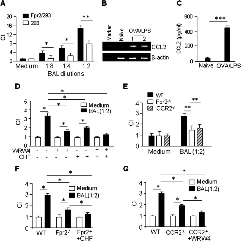
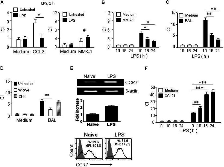
Chemoattractant receptors regulate leukocyte accumulation at sites of inflammation. In allergic airway inflammation, although a chemokine receptor CCR2 was implicated in mediating monocyte-derived dendritic cell (DC) recruitment into the lung, we previously also discovered reduced accumulation of DCs in the inflamed lung in mice deficient in formylpeptide receptor Fpr2 (Fpr2(-/-)). We therefore investigated the role of Fpr2 in the trafficking of monocyte-derived DCs in allergic airway inflammation in cooperation with CCR2. We report that in allergic airway inflammation, CCR2 mediated the recruitment of monocyte-derived DCs to the perivascular region, and Fpr2 was required for further migration of the cells into the bronchiolar area. We additionally found that the bronchoalveolar lavage liquid from mice with airway inflammation contained both the CCR2 ligand CCL2 and an Fpr2 agonist CRAMP. Furthermore, similar to Fpr2(-/-) mice, in the inflamed airway of CRAMP(-/-) mice, DC trafficking into the peribronchiolar areas was diminished. Our study demonstrates that the interaction of CCR2 and Fpr2 with their endogenous ligands sequentially mediates the trafficking of DCs within the inflamed lung.

Keywords: CCR2; Chemokines; Chemotaxis; Dendritic Cells; Fpr2; Lung; Monocytes; Peribronchiole Region; Perivascular Region.


Figures

FIGURE 1.
FIGURE 1.
Reduced accumulation of CD11c+MHC II+CD11b+ DCs in the inflamed airway in Fpr2−/− mice. A and B, accumulation of CD11c+ MHC II+ cells in the inflamed lung. The results are expressed as the means ± S.E. n = 5 mice/group. ***, p < 0.001. C and D, the frequency of CD11b+ cells in the CD11c+ MHC II+ cell population. The results are expressed as the means ± S.E. n = 5 mice/group. **, p < 0.01; ***, p < 0.001. Na, naïve; OL, OVA/LPS.
FIGURE 2.
FIGURE 2.
Reduced accumulation of monocyte-derived Ly6C+ DCs in the lung of Fpr2−/− mice with allergic airway inflammation. A and B, accumulation of CD11c+ cells in the lung of mice with allergic airway inflammation. The results are expressed as the means ± S.E. n = 5 mice/group. **, p < 0.01. C and D, the percentage of CD11b+Ly6C+ cells in the CD11c+ cell population. The results are expressed as the means ± S.E. n = 5 mice/group. ***, p < 0.001. E, cumulative frequency of CD11b+Ly6C+CD11c+ cells in the lung. The results are expressed as the means ± S.E. n = 5 mice/group. **, p < 0.01; ***, p < 0.001. N, naïve; OL, OVA/LPS.
FIGURE 3.
FIGURE 3.
Monocyte-derived DC populations in the lung and blood of Fpr2−/− mice with allergic airway inflammation. A, frequency of CD11c+CCR2+ cells in the Ly6C+ population in the inflamed lung of mice. B, cumulative results for CCR2+, CCR5+, and CCR6 + cells in the CD11c+Ly6C+ cell population in the inflamed lung. The results are expressed as the means ± S.E. n = 5 mice/group. The values are calculated as follows: percentage of CCR2+CD11c+Ly6C+ cell population = percentage of Ly6C+ cell population × percentage of CCR2+CD11c+/100. *, p < 0.05. C, accumulation of CD11b+ cells in the circulation. D, cumulative results of CCR2+ CD11b+Ly6C+ cell frequencies in the blood. The results are expressed as the means ± S.E. n = 5 mice/group. OL, OVA/LPS. **, p < 0.01; ***, p < 0.001.
FIGURE 4.
FIGURE 4.
Reduced inflammatory cell infiltration in the peribronchiole regions of the inflamed lung of Fpr2−/− mice and reduced recruitment of Ly6C+ cells into the inflamed lung in CCL2−/− mice. A, inflammatory cell infiltration in the peribronchiole regions in the inflamed lung. A, alveolus; B, bronchiole; V, blood vessel. Scale bar, 100 μm. The results from a representative experiment of four performed are shown. B, cumulative disease scores of perivascular regions in the lung. C, cumulative disease scores of the peribronchiole regions in the lung. The results are expressed as the means ± S.E. obtained from 45 blood vessels and 40 bronchioles of eight mice/group. ***, p < 0.001. D, reduced inflammatory cell infiltration in the lung of CCL2−/− mice. Scale bar, 100 μm. The results from a representative experiment of four performed are shown. E, absence of recruitment of Ly6C+ cells into the inflamed lung in CCL2−/− mice. CCL2−/− mice were immunized with OVA and aerosol-challenged with OVA/LPS. CD11c+Ly6C+ cell population and the percentage of CCR2+CD11c+ cells in the Ly6C+ population were analyzed with FACS. The data from a representative experiment of four performed are shown. The results are expressed as the means ± S.E., n = 5 mice/group.
FIGURE 5.
FIGURE 5.
Reduced accumulation of CD11c+CD11b+ myeloid cells in the lung of Fpr2−/− mice with allergic airway inflammation. A, reduced CD11c+CD11b+ myeloid cell infiltration in the inflamed lung of Fpr2−/− mice. The mice were immunized with OVA and aerosol-challenged with OVA/LPS. Frozen lung sections were stained with hamster anti-mouse CD11c and CD11b antibodies followed by biotinylated anti-Ig antibodies and streptavidin-PE (red) or streptavidin-FITC (green). The nuclei were revealed by DAPI (blue). Hamster IgG was used as an isotype control. Scale bar, 50 μm. The results from a representative experiment of three performed are shown. B, fluorescence intensity of CD11c+CD11b+ cells in the lung. The mice used were 8–12-week-old male littermates. ***, p < 0.001, significantly reduced CD11c+CD11b+ cells in the inflamed lung of Fpr2−/− mice as compared with WT mice. CD11c+CD11b+ cells in the lung in mediastinal LN were identified by immunofluorescence. Frozen sections were stained with anti-mouse CD11c (BD Biosciences) and anti-mouse CD11b (BD Biosciences) followed by biotinylated anti-Ig Abs (BD Biosciences) with streptavidin-PE or streptavidin-FITC and DAPI (Invitrogen) or PE-anti-rabbit IgG antibody. Hamster IgG, rabbit IgG, or rat IgG2b was used as an isotype control. OL, OVA/LPS.
FIGURE 6.
FIGURE 6.
Restoration by adoptive transfer of WT mouse BM cells. A, cell infiltration in the lung tissues surrounding the inflamed bronchioles in chimeric mice. A, alveolus; B, bronchiole; V, blood vessel. Scale bar, 100 μm. B, cumulative disease scores of the perivacular and bronchiole regions in the inflamed lung. The results are expressed as the means ± S.E. from 20 blood vessels and 25 bronchioles of 5 mice/group. *, p < 0.05; ***, p < 0.001, significantly reduced scores in the perivascular regions of the inflamed lung in Fpr2−/− mice as compared with WT mice and significantly restored scores in the peribronchiole regions in the lung of Fpr2−/− mice receiving transfer of WT BM cells as compared with Fpr2−/− mice without BM transfer. C, restoration of CD11c+Ly6C+ cells infiltrating the inflamed lung tissues of chimera mice. The results from a representative experiment of four performed are shown.
FIGURE 7.
FIGURE 7.
Fpr2 and CCR2 agonists contained in BAL from WT mice with allergic airway inflammation. A, parental 293 cells and 293 cells transfected with Fpr2 (Fpr2/293) were measured for migration induced by different concentrations of BAL from WT mice with airway inflammation. *, p < 0.05; **, p < 0.01. B, CCL2 mRNA expression in lung tissues. C, CCL2 in BAL. The results are expressed as the means ± S.E. n = 5 mice/group. ***, p < 0.001. D, inhibition of BAL-induced DC chemotaxis by CCR2 or Fpr2 antagonist. *, p < 0.05. E, reduced chemotaxis of DCs from Fpr2−/− and CCR2−/− mice in response to BAL. **, p < 0.01. F, CCR2 antagonist inhibits Fpr2−/− cell migration to BAL. *, p < 0.05. G, inhibition of CCR2−/− DC migration to BAL by Fpr2 antagonist. *, p < 0.05. CI, chemotaxis index.
FIGURE 8.
FIGURE 8.
Chemotactic activity of CRAMP in the BAL of mice with allergic airway inflammation. A, CRAMP expression in the lung. Densitometry analysis shows significant increase of CRAMP in the inflamed lung of WT mice (*, p < 0.01). B, reduction of the chemotactic activity of BAL for DCs by anti-CRAMP antibody. The results are expressed as the means ± S.E. *, p < 0.05; **, p < 0.01. C, reduction of the chemotactic activity of BAL from CRAMP−/− mice with allergic airway inflammation. *, p < 0.05. D, parental 293 cells and 293 cells transfected with Fpr2 (Fpr2/293) were measured for migration induced by the BAL from CRAMP−/− mice with airway inflammation. **, p < 0.01. CI, chemotaxis index.
FIGURE 9.
FIGURE 9.
BAL-induced chemotaxis of WT mouse DCs activated with LPS (100 ng/ml). A, down-regulation of CCR2 function and increased Fpr2 function in DCs 1 h after LPS stimulation. *, p < 0.05; #, p < 0.05. B, time-dependent Fpr2 function after LPS stimulation. *, p < 0.05. C, time-dependent migration of DCs from WT mice in response to BAL (1:2) after LPS stimulation. **, p < 0.01. D, failure of CCR2 antagonist to inhibit the migration of LPS-treated DCs in response to BAL. **, p < 0.01. E, CCR7 expression in DCs after LPS stimulation. Top panel, CCR7 mRNA expression. Middle panel, fold increase of CCR7 mRNA expression after LPS stimulation. Bottom panel, CCR7 protein expression. MFI, Mean Fluorescence Intensity. F, increased CCR7 function in inflammatory DCs after LPS stimulation. **, p < 0.01; ***, p < 0.001. CI, chemotaxis index.

References

    1. Holt P. G., Upham J. W. (2004) The role of dendritic cells in asthma. Curr. Opin. Allergy Clin. Immunol. 4, 39–44 - PubMed
    1. Lambrecht B. N., Hammad H. (2009) Biology of lung dendritic cells at the origin of asthma. Immunity 31, 412–424 - PubMed
    1. GeurtsvanKessel C. H., Lambrecht B. N. (2008) Division of labor between dendritic cell subsets of the lung. Mucosal Immunol. 1, 442–450 - PubMed
    1. Domínguez P. M., Ardavín C. (2010) Differentiation and function of mouse monocyte-derived dendritic cells in steady state and inflammation. Immunol. Rev. 234, 90–104 - PubMed
    1. Nakano H., Lin K. L., Yanagita M., Charbonneau C., Cook D. N., Kakiuchi T., Gunn M. D. (2009) Blood-derived inflammatory dendritic cells in lymph nodes stimulate acute T helper type 1 immune responses. Nat. Immunol. 10, 394–402 - PMC - PubMed

Publication types

MeSH terms