. 2013 May;34(5):681-90.
doi: 10.1038/aps.2013.22. Epub 2013 Apr 22.

NVP-BEZ235, a novel dual PI3K/mTOR inhibitor, enhances the radiosensitivity of human glioma stem cells in vitro

Affiliations

NVP-BEZ235, a novel dual PI3K/mTOR inhibitor, enhances the radiosensitivity of human glioma stem cells in vitro

Wen-juan Wang et al. Acta Pharmacol Sin. 2013 May.

Abstract

Aim: NVP-BEZ235 is a novel dual PI3K/mTOR inhibitor and shows dramatic effects on gliomas. The aim of this study was to investigate the effects of NVP-BEZ235 on the radiosensitivity and autophagy of glioma stem cells (GSCs) in vitro.

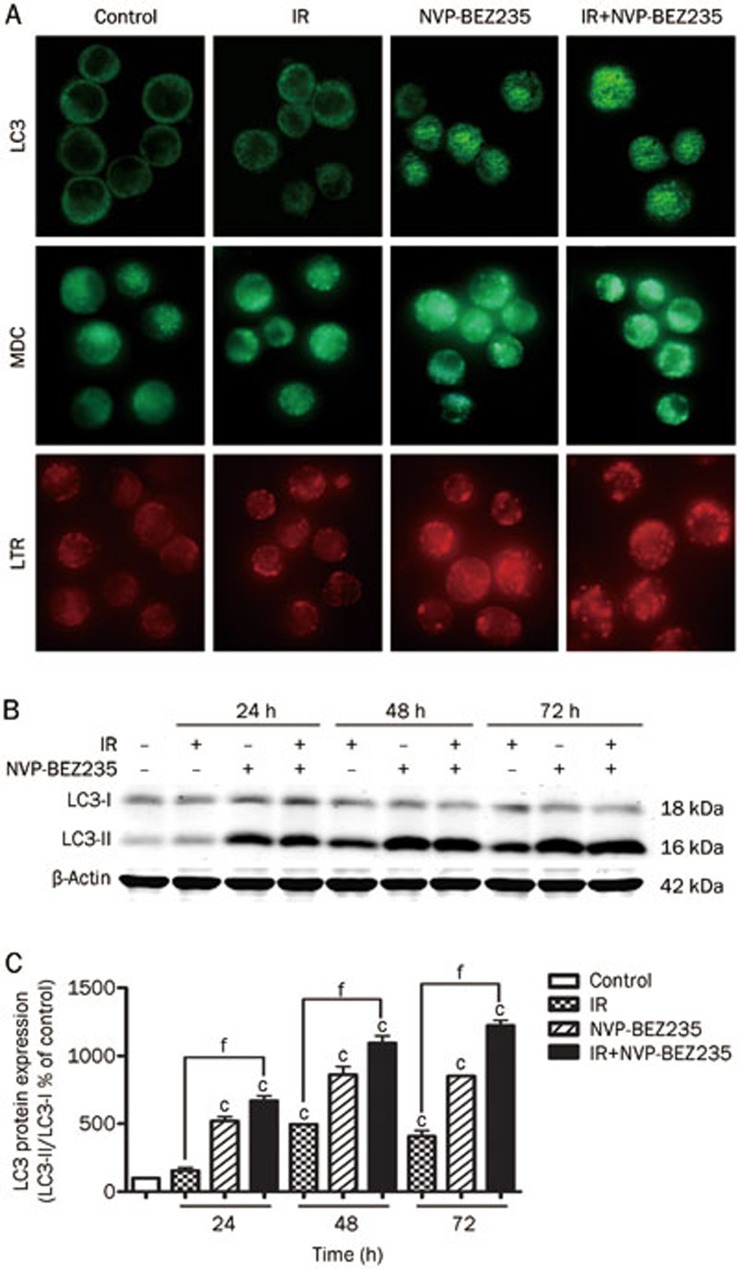
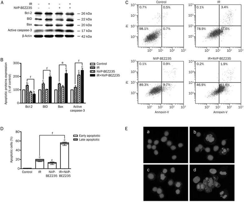
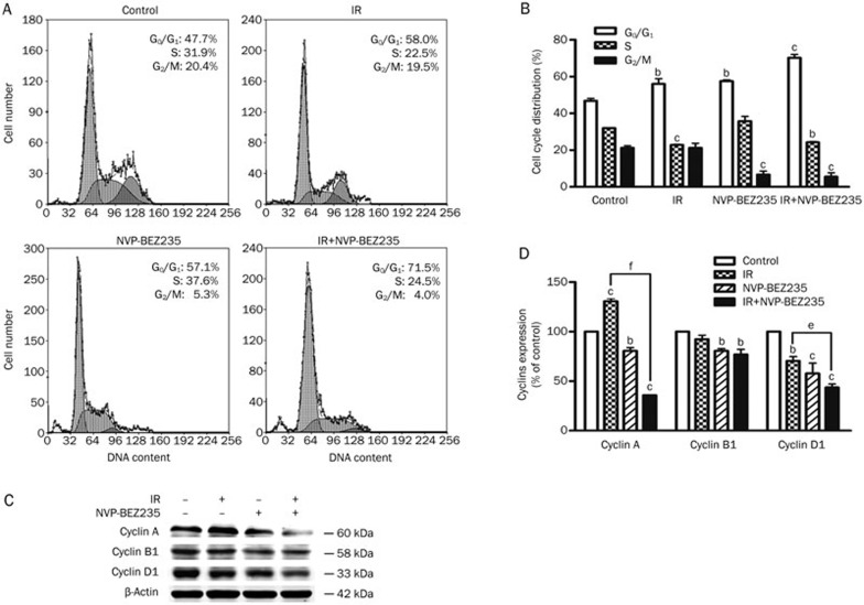
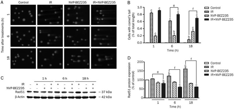
Methods: Human GSCs (SU-2) were tested. The cell viability and survival from ionizing radiation (IR) were evaluated using MTT and clonogenic survival assay, respectively. Immunofluorescence assays were used to identify the formation of autophagosomes. The apoptotic cells were quantified with annexin V-FITC/PI staining and flow cytometry, and observed using Hoechst 33258 staining and fluorescence microscope. Western blot analysis was used to analyze the expression levels of proteins. Cell cycle status was determined by measuring DNA content after staining with PI. DNA repair in the cells was assessed using a comet assay.

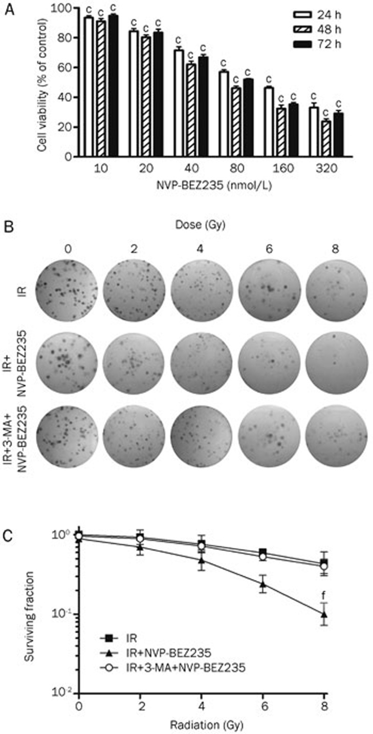
Results: Treatment of SU-2 cells with NVP-BEZ235 (10-320 nmol/L) alone suppressed the cell growth in a concentration-dependent manner. A low concentration of NVP-BEZ235 (10 nmol/L) significantly increased the radiation sensitivity of SU-2 cells, which could be blocked by co-treatment with 3-MA (50 μmol/L). In NVP-BEZ235-treated SU-2 cells, more punctate patterns of microtubule-associated protein LC3 immunoreactivity was observed, and the level of membrane-bound LC3-II was significantly increased. A combination of IR with NVP-BEZ235 significantly increased the apoptosis of SU-2 cells, as shown in the increased levels of BID, Bax, and active caspase-3, and decreased level of Bcl-2. Furthermore, the combination of IR with NVP-BEZ235 led to G1 cell cycle arrest. Moreover, NVP-BEZ235 significantly attenuated the repair of IR-induced DNA damage as reflected by the tail length of the comet.

Conclusion: NVP-BEZ235 increases the radiosensitivity of GSCs in vitro by activating autophagy that is associated with synergistic increase of apoptosis and cell-cycle arrest and decrease of DNA repair capacity.


Figures

Figure 1
Figure 1
NVP-BEZ235 inhibits GSC proliferation and enhances radiosensitivity. (A) SU-2 cells were treated with increasing concentrations of NVP-BEZ235 for 24, 48, or 72 h and subjected to MTT assay. The plot depicts the percentage growth of the NVP-BEZ235-treated cells compared with that of the untreated control cells (the viability of the untreated control cells was considered as 100%). (B) Clonogenic survival assay in SU-2 cells treated with radiation alone (0 Gy to 8 Gy), radiation plus NVP-BEZ235 (10 nmol/L), or radiation plus 3-MA (50 μmol/L) and NVP-BEZ235 (10 nmol/L). (C) Clonogenic survival curves for (B). Mean±SD. n=3. cP<0.01 compared with the control group, fP<0.01 compared with the IR-alone group.
Figure 2
Figure 2
NVP-BEZ235-mediated radiosensitization increases autophagy induction. (A) SU-2 cells were treated with NVP-BEZ235 (10 nmol/L) for 12 h and then harvested 24 h after treatment with IR (8 Gy), and analyzed for LC3 expression (top) or the formation of autophagolysosomes by using MDC (middle) and LTR (bottom) by immunofluorescence analysis (600×magnification). (B) The cells were treated with IR and NVP-BEZ235 and analyzed for LC3 and β-actin expression by Western blot analysis. β-Actin was used as a loading control. (C) Quantification of LC3 protein expression in SU-2 cells treated by indicated treatments after normalization with β-actin levels. Mean±SD. n=3. cP<0.01 compared with the control group. fP<0.01 compared with the IR-alone group.
Figure 3
Figure 3
Induction of GSC apoptosis by co-treatment with IR and NVP-BEZ235. (A) SU-2 cells were collected after 48 h of treatment with IR (8 Gy) and NVP-BEZ235 (10 nmol/L), protein lysates were resolved by SDS-PAGE, and immunoblotted with antibodies for Bcl-2, BID, Bax, and active caspase-3. Equal protein loading was confirmed by blotting for β-actin. (B)Quantification of apoptotic proteins expression in SU-2 cells treated by IR and NVP-BEZ235 treatments after normalization with β-actin levels. (C) After treatment with IR and NVP-BEZ235, the cells were stained with annexin V-FITC/PI. The co-treated group demonstrated a dramatic increase in the percentage of apoptotic cells compared with the groups that were untreated (control), treated with IR-alone, or treated with NVP-BEZ235-alone. (D) The quantification of the percentage of apoptotic cells result from (C). (E) The SU-2 cell nuclei were stained with Hoechst 33258 to detect apoptosis morphologically (400×magnification), a) control group; b) IR-alone group; c) NVP-BEZ235-alone group; d) co-treated group. Microphotographs are shown as representative results from three independent experiments. Mean±SD. n=3. bP<0.05, cP<0.01 compared with the control group. eP<0.05, fP<0.01 compared with the IR-alone group.
Figure 4
Figure 4
G1 phase blockade induced by IR and NVP-BEZ235 treatments. (A) SU-2 cells were treated with IR (8 Gy) and NVP-BEZ235 (10 nmol/L) and harvested 48 h after IR. Cellular DNA content was determined by staining with a hypotonic PI solution. (B) Quantification of cell-cycle distribution in SU-2 cells. (C) Western blot analysis of cyclins in GSCs. SU-2 cells were harvested 48 h after IR. (D) The quantitation of protein expression in response to IR and NVP-BEZ235 treatments after normalization with β-actin levels. Mean±SD. n=3. bP<0.05, cP<0.01 compared with the control group. eP<0.05, fP<0.01 compared with the IR-alone group.
Figure 5
Figure 5
NVP-BEZ235 impairs the repair of radiation-induced DNA damage in GSCs. (A) GSCs were treated with IR (8 Gy) and NVP-BEZ235 (10 nmol/L) and harvested at 1, 6, and 18 h after IR. The alkaline comet assay was performed to determine treatment-induced DNA damage. Representative images of SU-2 cells at indicated time points after various treatments are shown (400×magnification). (B) The quantification of the percentage of cells with comet tails at different time points. (C) Cell lysates were analyzed by Western blot analysis with the indicated antibodies. (D) The relative abundance of each band to β-actin levels was quantified, and the control levels were set at 100%. Mean±SD. n=3. bP<0.05, cP<0.01 compared with the control group. eP<0.05, fP<0.01 compared with the IR-alone group.

Similar articles

Cited by

References

    1. Tunici P, Irvin D, Liu G, Yuan X, Zhaohui Z, Ng H, et al. Brain tumor stem cells: new targets for clinical treatments. Neurosurg Focus. 2006;20:E27. - PubMed
    1. Gupta T, Nair V, Paul SN, Kannan S, Moiyadi A, Epari S, et al. Can irradiation of potential cancer stem-cell niche in the subventricular zone influence survival in patients with newly diagnosed glioblastoma. J Neurooncol. 2012;109:195–203. - PubMed
    1. Singh SK, Hawkins C, Clarke ID, Squire JA, Bayani J, Hide T, et al. Identification of human brain tumour initiating cells. Nature. 2004;432:396–401. - PubMed
    1. Qiu B, Zhang D, Tao J, Wu A, Wang Y. A simplified and modified procedure to culture brain glioma stem cells from clinical specimens. Oncol Lett. 2012;3:50–4. - PMC - PubMed
    1. Jamal M, Rath BH, Tsang PS, Camphausen K, Tofilon PJ. The brain microenvironment preferentially enhances the radioresistance of CD133 (+) glioblastoma stem-like cells. Neoplasia. 2012;14:150–8. - PMC - PubMed

Publication types

MeSH terms