. 2013 May 15;32(10):1440-50.
doi: 10.1038/emboj.2013.86. Epub 2013 Apr 19.

Species-specific detection of the antiviral small-molecule compound CMA by STING

Affiliations

Species-specific detection of the antiviral small-molecule compound CMA by STING

Taner Cavlar et al. EMBO J. .

Abstract

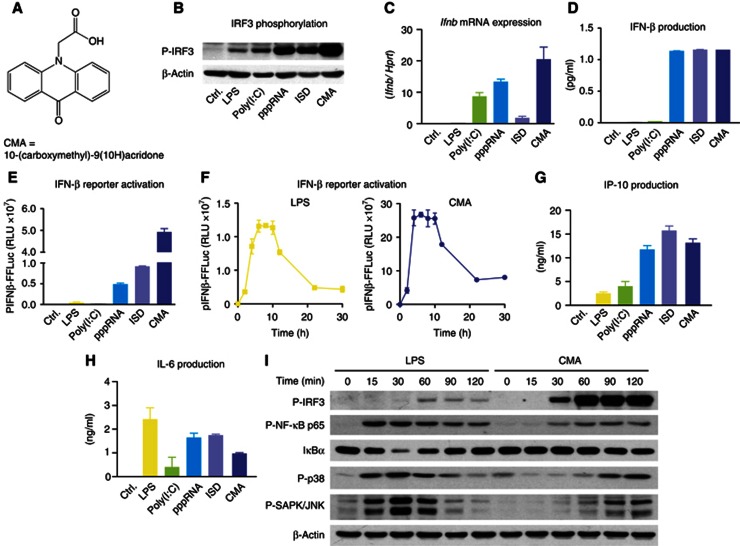
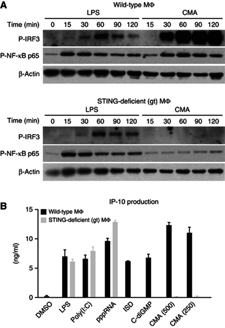
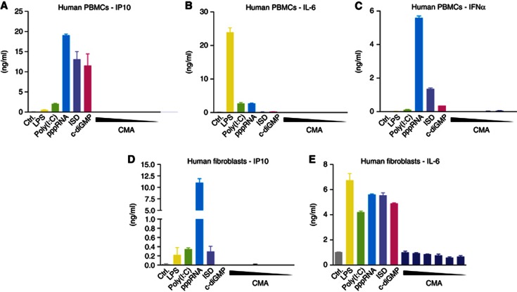
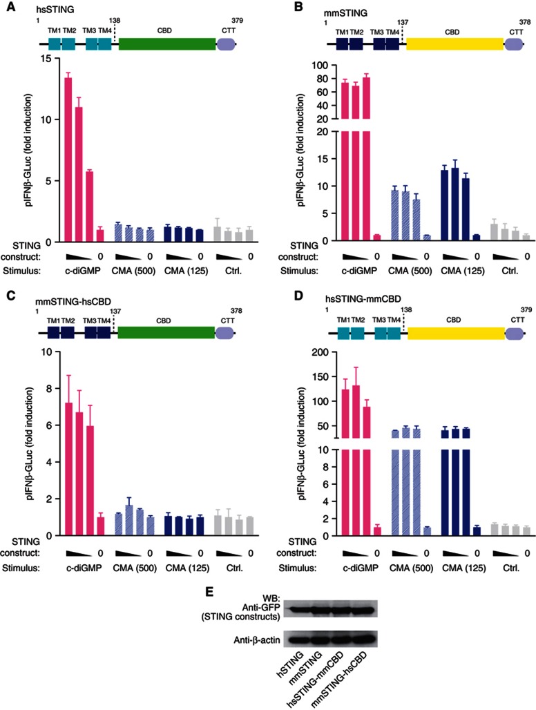
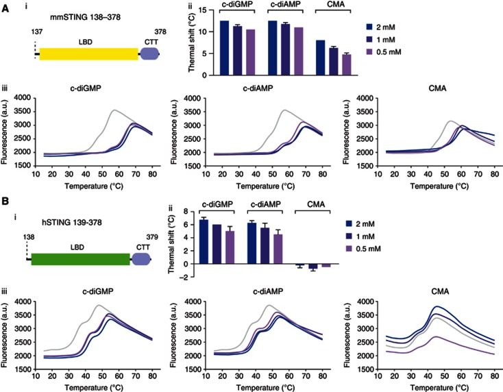
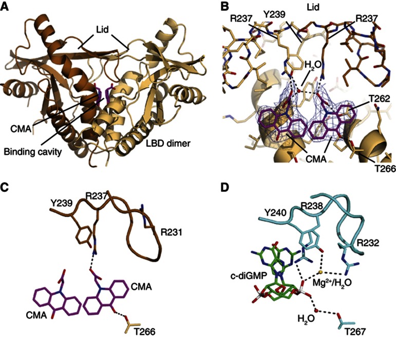
Extensive research on antiviral small molecules starting in the early 1970s has led to the identification of 10-carboxymethyl-9-acridanone (CMA) as a potent type I interferon (IFN) inducer. Up to date, the mode of action of this antiviral molecule has remained elusive. Here we demonstrate that CMA mediates a cell-intrinsic type I IFN response, depending on the ER-resident protein STING. CMA directly binds to STING and triggers a strong antiviral response through the TBK1/IRF3 route. Interestingly, while CMA displays extraordinary activity in phosphorylating IRF3 in the murine system, CMA fails to activate human cells that are otherwise responsive to STING ligands. This failure to activate human STING can be ascribed to its inability to bind to the C-terminal ligand-binding domain of human STING. Crystallographic studies show that two CMA molecules bind to the central Cyclic diguanylate (c-diGMP)-binding pocket of the STING dimer and fold the lid region in a fashion similar, but partially distinct, to c-diGMP. Altogether, these results provide novel insight into ligand-sensing properties of STING and, furthermore, unravel unexpected species-specific differences of this innate sensor.


Conflict of interest statement

The authors declare that they have no conflict of interest.

Figures

Figure 1
Figure 1
CMA strongly induces type I IFN in primary mouse macrophages. (A) The chemical structure of CMA is depicted. (B–E and G–H) Bone marrow-derived macrophages were transfected with poly(I:C), pppRNA and ISD, or stimulated with LPS or CMA (500 μg/ml). (B) After 2 h, cells were collected and subjected to SDS–PAGE, and western blotting for phospho-IRF3 (P-IRF3) was performed. (C) Four hours after stimulation, transcription of the IFNβ gene (Ifnb) was assessed by quantitative RT–PCR, with normalization to HPRT1. (D) Eighteen hours after stimulation, IFNβ was measured in the supernatants by enzyme-linked immunosorbent assay (ELISA). (E) pIFNβ-firefly-luciferase (pIFNβ-FFLuc) macrophages were stimulated as indicated. After 18 h, cells were lysed with passive lysis buffer, and FFLuc activity was measured in the lysates. (F) pIFNβ-FFLuc macrophages were stimulated with LPS or CMA (500 μg/ml). Luciferase activity was assessed at the indicated time points (in hours). (G,H) Eighteen hours after stimulation, IP-10 (G) and IL-6 (H) were measured in the supernatants by ELISA. (I) Bone marrow-derived macrophages were stimulated with LPS or CMA (500 μg/ml). Total protein was collected at indicated time points (in minutes) after stimulation and was assessed for P-IRF3, phospho-NF-κB-p65 (P-NF-κB p65), IkBα, phospho-p38 (P-p38) or phospho-SAPK/JNK (P-SAPK/JNK). Representative results out of three independent experiments are depicted. Source data for this figure is available on the online supplementary information page.
Figure 2
Figure 2
Loss of STING leads to complete abrogation of CMA-induced cytokine production. (A) Bone marrow-derived wild-type and STING-deficient macrophages were stimulated with LPS and CMA (500 μg/ml). Phospho-IRF3 (P-IRF3) and phospho-NF-κB-p65 (P-NF-κB p65) were assessed at indicated time points (in minutes) after stimulation. (B) Bone marrow-derived wild-type and STING-deficient macrophages were transfected with poly(I:C), pppRNA, ISD and c-diGMP, or stimulated with LPS and CMA (500 or 250 μg/ml) in duplicates. Supernatants were collected after 18 h, and IP-10 levels were measured by enzyme-linked immunosorbent assay. Representative results out of two independent experiments are depicted, whereas data are presented as mean values+s.e.m. Source data for this figure is available on the online supplementary information page.
Figure 3
Figure 3
Human cells do not respond to CMA. (AE) Primary human PBMCs and fibroblasts were transfected with poly(I:C), pppRNA, ISD and c-diGMP, or stimulated with LPS and decreasing concentrations of CMA (4000—125 μg/ml in twofold dilutions). IP-10, IL-6 and IFNα levels in the supernatants of stimulated PBMCs (AC) and fibroblasts (D,E) were determined by enzyme-linked immunosorbent assay. Representative results out of three independent experiments are depicted.
Figure 4
Figure 4
Species-specificity CMA activity is determined by the C-terminal LBD of STING. (A–D) 293T cells were transiently transfected with the indicated STING constructs (25, 12.5, 6.25 and 0 ng), whereas 12.5 ng of pIFNα-GLuc reporter plasmid were included. For titrations, an empty pCI vector served as a stuffer to obtain 200 ng total plasmid DNA. After 24 h, cells were transfected with c-diGMP or stimulated with CMA (500 and 125 μg/ml). Luciferase activity was measured after an additional period of 24 h in the supernatant, and data were normalized to the condition without STING overexpression. Plasmids coding for full-length hSTING (A), mmSTING (B), mmSTING(1-137)-hSTING(139-379) (C) and hSTING(1-138)-mmSTING(138-378) (D) were tested. (E) Expression of the above described constructs was studied in 293T cells 24 h after transfection (200 ng per 96-well plate) using western blot, whereas β-actin served as a loading control. Representative results out of three independent experiments are depicted. Source data for this figure is available on the online supplementary information page.
Figure 5
Figure 5
DSF implicates direct binding of CMA to murine, but not to human STING. (A,B) The interaction of STING with c-diGMP, c-diAMP and CMA were analysed by thermal shift assay. Purified murine STING (A) and human STING (B) were tested with different concentrations of c-diGMP/c-diAMP/CMA; (i) Schematic views of the protein domains used for binding studies are shown; (ii) thermal shifts of (iii) fluorescence intensity versus temperature are shown. Representative results out of two independent experiments are depicted for the temperature curves (ii), whereas mean values+s.e.m. out of two independent experiments are depicted for the thermal shift graphs (iii).
Figure 6
Figure 6
Structural basis for CMA recognition. (A) Ribbon model of the mouse STING dimer (light and dark brown) with highlighted secondary structure. The two bound CMA molecules are shown as magenta stick models. (B) Close-up view of the CMA-binding site with superimposed 2mFo-DFc electron density (blue; contoured at 1.4). One STING protomer is shown in light brown. For the other protomer (brown), only the lid is displayed. Folding of the lid via hydrogen bonds to Arg237 and Tyr239 suggests how CMA activates mouse STING. (C,D) Side-by-side comparison of CMA bound to mouse STING (C) and c-diGMP bound to human STING (D) showing selected interactions. CMA folds the lid differently from c-diGMP, due to steric clash with Arg231 (human Arg232), which binds c-diGMP via a magnesium ion/water molecule.

References

    1. Adams PD, Afonine PV, Bunkoczi G, Chen VB, Davis IW, Echols N, Headd JJ, Hung LW, Kapral GJ, Grosse-Kunstleve RW, McCoy AJ, Moriarty NW, Oeffner R, Read RJ, Richardson DC, Richardson JS, Terwilliger TC, Zwart PH (2010) PHENIX: a comprehensive Python-based system for macromolecular structure solution. Acta Crystallogr D Biol Crystallogr 66: 213–221 - PMC - PubMed
    1. Ahn J, Gutman D, Saijo S, Barber GN (2012) STING manifests self DNA-dependent inflammatory disease. Proc Natl Acad Sci USA 109: 19386–19391 - PMC - PubMed
    1. Arnold K, Bordoli L, Kopp J, Schwede T (2006) The SWISS-MODEL workspace: a web-based environment for protein structure homology modelling. Bioinformatics 22: 195–201 - PubMed
    1. Aslanidis C, de Jong PJ (1990) Ligation-independent cloning of PCR products (LIC-PCR). Nucleic Acids Res 18: 6069–6074 - PMC - PubMed
    1. Berger Rentsch M, Zimmer G (2011) A vesicular stomatitis virus replicon-based bioassay for the rapid and sensitive determination of multi-species type I interferon. PLoS One 6: e25858. - PMC - PubMed

Publication types

MeSH terms

Associated data