. 2013 May;20(5):547-54.
doi: 10.1038/nsmb.2565. Epub 2013 Apr 21.

HDAC-mediated suppression of histone turnover promotes epigenetic stability of heterochromatin

Affiliations

HDAC-mediated suppression of histone turnover promotes epigenetic stability of heterochromatin

Ozan Aygün et al. Nat Struct Mol Biol. 2013 May.

Abstract

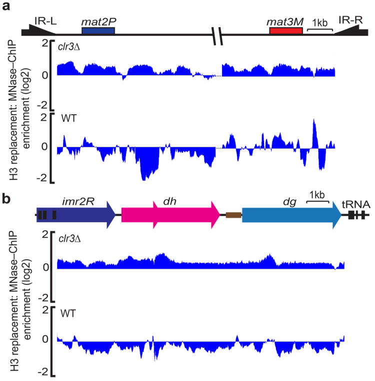
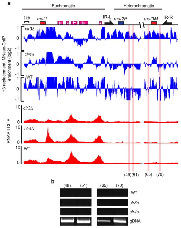
Heterochromatin causes epigenetic repression that can be transmitted through multiple cell divisions. However, the mechanisms underlying silencing and stability of heterochromatin are not fully understood. We show that heterochromatin differs from euchromatin in histone turnover and identify histone deacetylase (HDAC) Clr3 as a factor required for inhibiting histone turnover across heterochromatin domains in Schizosaccharomyces pombe. Loss of RNA-interference factors, Clr4 methyltransferase or HP1 proteins involved in HDAC localization causes increased histone turnover across pericentromeric domains. Clr3 also affects histone turnover at the silent mating-type region, where it can be recruited by alternative mechanisms acting in parallel to H3K9me-HP1. Notably, the JmjC-domain protein Epe1 promotes histone exchange, and loss of Epe1 suppresses both histone turnover and defects in heterochromatic silencing. Our results suggest that heterochromatic-silencing factors preclude histone turnover to promote silencing and inheritance of repressive chromatin.


Figures

Figure 1
Figure 1. Differential patterns of histone turnover define heterochromatin and euchromatin domains
(a) Schematic depiction of the experimental design used to investigate histone H3 replacement. (b) Western blot (WB) analysis of the induction of H3–FLAG fusion protein expression upon a switch in carbon source from glucose (glu) to sucrose (suc) for 2h. Ponceau S staining is shown as loading control. (c) FACS analysis of DNA replication arrest and synchronization of cells. N and 2N refer to DNA content before and after DNA replication, respectively. (d) Distribution of H3 replacement across a pericentromeric domain and the adjacent euchromatic region. H3 replacement values, as measured by MNase–ChIP–chip (ChIP vs Input), are plotted in alignment with the map of the right pericentromeric region of cen2 and the neighboring euchromatic region (top). Heterochromatin domain containing the inner centromeric repeat (imr2R) as well as the dg and dh repeat elements is indicated. Vertical lines indicate tRNAs. Nucleosome occupancy (measured by MNase hypersensitivity) is shown for comparison (bottom). (e) Distribution of H3 replacement and nucleosome occupancy across the mating type region illustrated shown as in d. Arrows mark the inverted repeats IR–L and IR–R that act as boundary elements. The dotted lines and gap between mat2P and mat3M represent the probes that correspond to the cenH region, which are omitted due to cross–hybridization with centromeric repeats. Since non–switching mat1M–Smt0 cells were used to perform experiments, signal mapping to mat3M may represent cross hybridization to the mat1M locus.
Figure 2
Figure 2. Clr4 and RNAi are required to suppress H3 replacement at centromeres but not at the silent mating–type locus
(a) H3 replacement across the right pericentromeric region of cen2 in clr4Δ, dcr1Δ or wild–type (WT) cells was measured by MNase–ChIP–chip method as described in Fig 1. (b) H3 replacement across the silent mating–type region in clr4Δ or dcr1Δ, measured and shown as in a.
Figure 3
Figure 3. HP1 proteins Chp2 and Swi6 cooperate to prevent nucleosome turnover across a pericentromeric loci
(a) Histone H3 replacement in swi6Δ chp2Δ, chp2Δ, swi6Δ, or wild–type (WT) cells was measured by MNase–ChIP–chip method as described in Fig.1 and plotted in alignment with the map of the right pericentromeric region of cen2. (b) Histone H3 turnover across the silent mating–type region. H3 replacement was measured and shown as in a.
Figure 4
Figure 4. Clr3 HDAC is required for suppression of histone H3 exchange across heterochromatin domains
Histone H3 replacement was measured across the silent mating type region (a) and pericentromeric heterochromatin (b) in clr3Δ or wild–type (WT) cells as described in Fig.1.
Figure 5
Figure 5. Increased H3 turnover in heterochromatin mutants is not solely due to changes in RNAPII transcription
(a) Histone H3 replacement (blue) was measured across the mating type locus using MNase–ChIP–on–Chip analysis as described in Fig 1. RNAPII occupancy (ChIP vs Input) was measured by ChIP–on–Chip in clr3Δ, clr4Δ or wild–type (WT) cells and plotted in alignment with the map (red). (b) RT–PCR analysis performed using total RNA samples isolated from clr3Δ, clr4Δ or wild–type (WT) cells. Genomic DNA (gDNA) was used as a control. The locations amplified by primer pairs 49, 51, 65 and 70 are highlighted with red shading (see online methods for primer references). Heterochromatin and euchromatin portions of the mating–type region are indicated at the top.
Figure 6
Figure 6. The JmjC domain–containing protein Epe1 promotes histone turnover across the pericentromeric regions
(a) Histone H3 replacement in Epe1 overexpressing (nmt1epe1), ago1Δ epe1Δ, ago1Δ, epe1Δ or wild–type (WT) cells was measured by the MNase–ChIP–chip method as described in Fig. 1 and plotted in alignment with the map of the right pericentromeric region of cen2. (b) RT–PCR analysis was used to measure expression at the cen–dh locus in epe1Δ, ago1Δ, ago1Δepe1Δ or wild–type (WT) cells. RNA isolated from epe1Δ, ago1Δ, ago1Δepe1Δ or wild–type (WT) cells was used to perform RT–PCR analysis with primer sets specific to centromeric dh repeats (cen–dh) or act1 loading control. (c) TBZ sensitivity of epe1Δ, ago1Δ, ago1Δepe1Δ or wild–type (WT) cells. Ten–fold serial dilutions of the indicated cultures were grown on rich medium (YEA) in the presence (10μg/ml) or absence of TBZ.
Figure 7
Figure 7. Clr3–dependent suppression of histone turnover correlates with epigenetic stability of heterochromatin
(a) ChIP analysis of Clr3 localization of at the silent mat region. Strains expressing Myc tagged Clr3 in KΔ∷ura4+ ura4–on or ura4–off state were used to perform ChIP. ChIP DNA was analyzed by semi–quantitative competitive PCR using primers that amplify both full–length KΔ∷ura4+ and endogenous mini–ura4 (ura4DSE) as internal control. The relative enrichments were determined by calculating the ratio of the band intensities of [ChIP KΔ∷ura4+ ÷ ChIP ura4DSE] ÷ [Input KΔ∷ura4+ ÷ Input ura4DSE]. Results were confirmed by quantitative real–time PCR (qPCR). Relative enrichment of KΔ∷ura4+ was normalized against untagged negative control and the mean enrichment is presented. Error bars represent standard error of the mean calculated from 3 independent biological replicates (n=3) (b) H3 replacement was measured in KΔ∷ura4+ ura4–on or ura4–off cells. The endogenous ura4+ was deleted in the strains used. (c) ChIP analysis of H3K9me2 levels at KΔ∷ura4+ ura4–on cells. Experiments were performed with the same strains used in a. H3K9me levels were confirmed by qPCR and the mean enrichment is presented. Error bars represent standard error of the mean calculated from 4 independent biological replicates (n=4).
Figure 8
Figure 8. Model showing effects of factors that impact epigenetic stability of heterochromatin
HDAC recruited by HP1 or other mechanisms are required to suppress nucleosome turnover and promote epigenetic stability of heterochromatin. In contrast, Epe1, which also associates with Swi6(HP1), stimulates histone exchange. The balance between these opposing activities that affect nucleosome turnover may underlie the epigenetic switch between ‘OFF’ and ‘ON’ states.

Similar articles

Cited by

References

    1. Felsenfeld G, Groudine M. Controlling the double helix. Nature. 2003;421:448–453. - PubMed
    1. Grewal SI, Jia S. Heterochromatin revisited. Nat Rev Genet. 2007;8:35–46. - PubMed
    1. Jenuwein T, Allis CD. Translating the histone code. Science. 2001;293:1074–1080. - PubMed
    1. Richards EJ, Elgin SC. Epigenetic codes for heterochromatin formation and silencing: rounding up the usual suspects. Cell. 2002;108:489–500. - PubMed
    1. Grunstein M. Histone acetylation in chromatin structure and transcription. Nature. 1997;389:349–352. - PubMed

Publication types

MeSH terms

Associated data