. 2014 Mar 27;33(13):1736-1742.
doi: 10.1038/onc.2013.126. Epub 2013 Apr 22.

MYC, a downstream target of BRD-NUT, is necessary and sufficient for the blockade of differentiation in NUT midline carcinoma

Affiliations

MYC, a downstream target of BRD-NUT, is necessary and sufficient for the blockade of differentiation in NUT midline carcinoma

Adlai R Grayson et al. Oncogene. .

Abstract

NUT midline carcinoma (NMC) is an aggressive type of squamous cell carcinoma that is defined by the presence of BRD-NUT fusion oncogenes, which encode chimeric proteins that block differentiation and maintain tumor growth. BRD-NUT oncoproteins contain two bromodomains whose binding to acetylated histones is required for the blockade of differentiation in NMC, but the mechanisms by which BRD-NUT act remain uncertain. Here, we provide evidence that MYC is a key downstream target of BRD4-NUT. Expression profiling of NMCs shows that the set of genes whose expression is maintained by BRD4-NUT is highly enriched for MYC upregulated genes, and MYC and BRD4-NUT protein expression is strongly correlated in primary NMCs. More directly, we find that BRD4-NUT associates with the MYC promoter and is required to maintain MYC expression in NMC cell lines. Moreover, both siRNA knockdown of MYC and a dominant-negative form of MYC, omomyc, induce differentiation of NMC cells. Conversely, differentiation of NMC cells induced by knockdown of BRD4-NUT is abrogated by enforced expression of MYC. Together, these findings suggest that MYC is a downstream target of BRD4-NUT that is required for maintenance of NMC cells in an undifferentiated, proliferative state. Our findings support a model in which dysregulation of MYC by BRD-NUT fusion proteins has a central role in the pathogenesis of NMC.


Figures

Figure 1
Figure 1. The MYC gene target signature and expression correlates with that of BRD-NUT
(a) Native BRD4-NUT-expressing NMC cells are enriched for the expression of MYC-upregulated genes. Previously published microarray data (5) from TC-797 and PER-403 NMC cell lines were ranked based on upregulation in control siRNA versus BRD4-NUT siRNA transfected cells and subjected to gene set enrichment analysis (GSEA) against curated gene sets within the Molecular Signatures Database (MSigDB)(6, 39). Shown are enrichment plots of genes shown previously to be upregulated by MYC (7) generated using GSEA software v2.0.9. (b) MYC is downregulated in differentiating NMC cells, in vivo. Anti-MYC and anti-NUT immunohistochemistry reveals staining of the same population of poorly differentiated tumor cells, and lack of staining in the same population of cells which have undergone squamous differentiation. NMC1 and NMC2 are BRD4-NUT NMCs excised from mediastinum and larynx, respectively. Immunohistochemical analysis for MYC and BRD4-NUT expression was performed as described (40, 41). Photomicrographs are of the same 400x magnification and are from the same field for each tumor.
Figure 2
Figure 2. The acetyl-histone binding of BRD4-NUT’s bromodomains are required for the expression of MYC in NMC
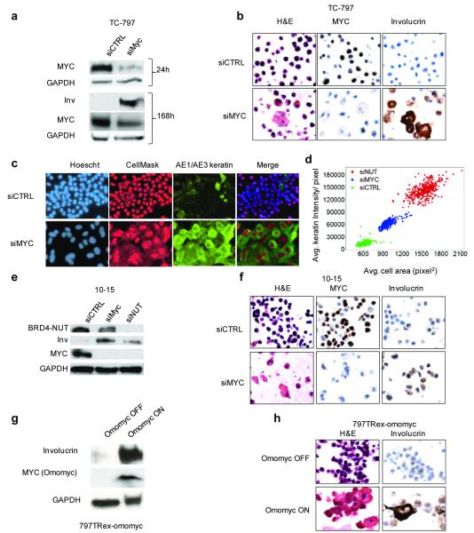
(a) BRD4-NUT enrichment at the MYC promoter region requires its bromodomains. Chromatin immunoprecipitation (ChIP) using 20 uL of anti-NUT (BX.1, whole rabbit serum) antibody in TC-797 cells treated with vehicle (DMSO) or the acetyl-histone mimic bromodomain inhibitor (BETi), JQ1 (500 nM (4)), for four hours. Samples were subjected to ChIP using the SimpleChIP Chromatin IP Kit from Cell Signaling Technologies (Danvers, MA). For each IP, 4 × 10⁷ cells were crosslinked with 1% formaldehyde for ten minutes at 37°C and nuclei were digested with micrococcal nuclease as per manufacturer instructions, with the exception that the “high salt” wash was at 200 mM NaCl. ChIPd DNA was eluted in 200 ul. 4 ul of eluted DNA was subjected to quantitative PCR (qPCR), in triplicate, using primers (Supplemental Table 1) designed to amplify the promoter-proximal region of MYC, and created based on distance from the transcriptional start site (TSS). qPCR was performed on a Bio-Rad iCycler in 96 well plate format with IQ SYBR Green supermix (Bio-Rad, Hercules, CA). Amplification curves and Ct values were generated using MyiQ Single-Color Real-Time software (Bio-Rad) using a range of inputs (10%, 0.1%, and 0.01%) to generate the standard curve. Results are depicted as percent input, and are representative of one of three separate experiments with standard deviations shown. Amino acids 800-1000 of NUT were used as immunogen to make polyclonal rabbit BX.1 antibody, using methods as described (42) (Chemicon, Temecula, CA). Primers used are listed in Supplemental Table 1. (b) BRD4-NUT maintains MYC transcription. mRNA levels, as measured by quantitative reverse-transcriptase PCR (qRT-PCR), of MYC, compared with those of the reference gene Ribosomal protein L13a (RPL13a), 24 h following siRNA-mediated knockdown of BRD4-NUT, or treatment with 500 nM JQ1. All qRT-PCR experiments are representative of triplicate qRT-PCRs from one of three independent experiments. Total RNA was harvested at the indicated time points using TRizol (Invitrogen, Carlsbad, CA) and the RNeasy Mini Kit (Qiagen, Valencia, CA). RNA (1 ug) was reversed transcribed into cDNA using the iScript cDNA synthesis kit (Bio-Rad, Hercules, CA). RT-qPCR was performed in triplicate on a Bio-Rad iCycler in 96 well plate format with IQ SYBR Green supermix (Bio-Rad) and 1 uL of cDNA template per reaction. Amplification curves and Ct values were generated using MyiQ Single-Color Real-Time software (Bio-Rad). Primers used are listed in Supplemental Table 1. (c) Immunoblot reveals that treatment of TC-797 NMC cells with JQ1 (500 nM) rapidly decreases MYC levels. Antibodies used were rabbit anti-MYC (1;1000, #9402, Cell Signaling Technology, Danvers, MA) and anti-GAPDH. siRNA was transfected into 2.5×10⁶ cells at a final concentration of 50 nM using the Nucleofector II (Lonza, Basel, Switzerland) electroporator, as described (3). (d) BRD4-NUT is necessary for maintenance of MYC protein expression. Immunoblot of lysates from TC-797 cells 24 h following transfection with control siRNA (siCTRL, siGENOME non-targeting siRNA #4, Dharmacon, Thermo Fisher Scientific, Waltham, MA) or siRNA directed at NUT (si3583, Dharmacon, sense, 5′ AAUUACCUUUGGAAGGAGCUAUU 3′, and anti-sense, 5′ UUUUAAUGGAAACCUUCCUCGAU 3′, oligonucleotides). Staining for BRD4-NUT was accomplished using AX.1 anti-NUT rabbit polyclonal antibody (1:500 (42)). (e) Schematic of BRD4-NUT, including known functional domains. (f-h) The acetyl-histone-binding bromodomains of BRD4-NUT are required for the blockade of differentiation and maintenance of MYC expression. (f) Immunophenotypic and morphologic (H&E) changes reveal differentiation in 797TRex-flag-BD12 cells expressing wild type tandem bromodomains 1 and 2 (wtBD12), but not when point mutations of key acetyl-histone binding residues are present in either or both bromodomains (N140A is the key acetyl-histone binding residue of bromodomain 1 (BD1), and N433A is that of bromodomain 2 (BD2)) of the tandem bromodomain construct (BD12), 120 h following induction. Differentiation is evidenced by morphologic (H&E) flattening and marked enlargement, and by increased expression of involucrin. (g) Immunoblot of lysates from 797TRex NMC cells 120h following tetracycline-induced expression (“OFF” is vehicle (ethanol) treated, and “ON” is tetracycline treated) of the dual flag-tagged tandem bromodomains (BD12) of BRD4, with and without (wt) point mutations that abrogate acetylhistone binding, demonstrates that only intact BD12 can induce differentiation, as evidenced by involucrin expression. Involucrin is a marker of squamous differentiation. Lysates 120 h after addition of tetracycline or ethanol were prepared with high-salt RIPA buffer (50 mM Tris, pH 8.0, containing 1% NP-40, 0.5% sodium deoxycholate, 0.1% SDS, 250 mM NaCl, and 5 mM EDTA) and separated by SDS-PAGE, electrophoretically transferred to Immobilon membranes (Millipore, Billerica, MA), and stained with mouse anti-GAPDH (1:5000, 6C5, Life Technologies/ Ambion, Grand Island, NY), mouse anti-flag (1:1000, Sigma-Aldrich, St. Louis, MO), and mouse anti-Involucrin (1:500, Sigma-Aldrich). Staining was developed using a chemiluminescent method (SuperSignal, West Pico; Pierce, Rockford, IL). The NMC cell line, TC-797, has been described (43). A TRex derivative of the NMC cell line, TC-797, containing a single FLP recombinase site (FRT), and multiple copies of tetracycline repressor gene (TR), to allow for the isogenic insertion of a desired tetracycline(tet)-inducible transgene, was created using the Flp-In T-Rex Core Kit and the pLenti6-TR plasmid, according the manufacturer’s instructions (Invitrogen). This derivative is termed 797TRex. Tetracycline-inducible 797TRex subclones were created according to the manufacturer’s instructions (Invitrogen, Carlsbad, CA) by cotransfection of TC-797 TRex cells (Invitrogen, Carlsbad, CA) with pCAGGS_FLPe and pcDNA5-Frt-TO plasmids encoding the following flag-epitope tagged polypeptides: BRD4 bromodomains 1 and 2 (BD12, residues 39-457 of BRD4); and BD12 with the point substitutions of the critical acetyl-histone-binding amino acids, N140A and N433A, alone or in combination(12). Clones were selected using Hygromycin (Invitrogen). Primers used to create these cDNAs and oligonucleotides used for site-directed mutagenesis (Stratagene/ Agilent Technologies, Inc., Santa Clara, CA) are provided in Supplemental Table 1. (h) Immunoblot revealing that induction of flag-wtBD12 results in decreased MYC protein levels.
Figure 3
Figure 3. MYC is necessary for the blockade of differentiation in NUT midline carcinoma
(a-b) Immunophenotypic and morphologic changes 120 h following siRNA transfection in TC-797 cells reveals differentiation in those cells subjected to siRNA knockdown of MYC (siMYC, siGENOME siRNA, Human MYC (4609), Dharmacon, Thermo Fisher Scientific) compared with control siRNA (siCTRL, siGENOME non-targeting siRNA #4). All photomicrographs are 400x and identical scale. Formalin-fixed, paraffin-embedded cell-blocks of cultured cells were prepared as described (3, 41). Sections were stained with hematoxylin and eosin or by immunohistochemistry (IHC), which was performed on 5 μm sections. Immunohistochemical stains performed using anti-MYC (rabbit monoclonal anti-human MYC antibody, Epitomics, Inc., Burlingame, CA, 1:500), and anti-involucrin (Sigma Aldrich, St. Louis, MO, 1:24,000) was as described (5). (c-d) High throughput analysis reveals that MYC is required to block differentiation of TC-797 cells. (c) Single well from a 384 well plate reveals morphologic (increased cell area, CellMask) and immunophenotypic (increased keratin intensity, green) changes in TC-797s consistent with differentiation 72 h following transfection with siMYC compared with siCTRL. Cells were transfected in 384 well format using 50 nM control siRNA (above), MYC siRNA (above), and NUT siRNA (above), as described (44). Cells were stained with HCS CellMask Red Stain (Life Technologies) to measure cell area, AE1/AE3 antibody (1:4, Dako, Carpinteria, CA) to measure keratin intensity, and nuclei were stained with Hoescht 33342 (4ug/ml, Molecular Probes) to quantitate cell numbers, as described. Imaging for cell size, keratin expression, and cell number was performed using the ImageXpress high-throughput microscope with MetaXpress software (Molecular Devices, Sunnyvale, CA) as described(44). Shown are representative images taken at 40x magnification. (d) Scatter plot of 384-well plate siRNA assay reveals differentiation of NMC cells transfected with siRNA to MYC and NUT. The differentiation phenotype is scored by an increase in cell area and keratin intensity. Each condition was performed in 384-well plate format, where 128 wells in triplicate were analyzed using MetaXpress with Multi Wavelength Cell Scoring for average cell area (pixels2) per well and average keratin fluorescence pixel intensity per well. Each well is represented by a dot graphed using Vortex Software (Dotmatics, Herts, UK). (e-f) Immunophenotypic and morphologic changes 120 h following siRNA knockdown of MYC demonstrate that MYC expression is also required to block differentiation in 10-15 cells, a primary cell culture obtained from the pleural fluid of a 42 year old man with BRD4-NUT NMC. Cells were initially grown in WIT media (5, 45), then maintained in standard media, as described for TC-797 cells (3). (g-h) The dominant-negative MYC polypeptide, omomyc, induces differentiation in NMC. Immunophenotypic and morphologic changes reveal differentiation in 797TRex-flag-omomyc cells 120 h following induction of omomyc expression compared with ethanol (EtOH) treated control cells.
Figure 4
Figure 4. MYC alone can block differentiation in NUT midline carcinoma
(a-c) MYC is sufficient to block differentiation in NMC. Tetracycline-induced (tet) or vehicle (ethanol) treated 797TRex-MYC cells were subjected to siRNA-mediated BRD4-NUT knockdown (MYC OFF siNUT, and MYC ON siNUT) versus control (MYC OFF, siCTRL or MYC ON, siCTRL). (a) Enlargement, stratification, flattening, and (a-b) involucrin expression, and (c) arrested proliferation are all markedly attenuated in cells subjected to BRD4-NUT knockdown and simultaneously induced to express MYC. All photomicrographs are 400x and identical scale. Three hundred cells were counted per sample for Ki-67 percentage. Standard deviations for triplicate counts are shown.

Similar articles

Cited by

References

    1. Bauer D, Mitchell C, Strait K, Lathan C, Stelow E, Lueer S, et al. Clinicopathologic Features and Long-Term Outcomes of NUT Midline Carcinoma. Clinical cancer research : an official journal of the American Association for Cancer Research. 2012;18(20):5773–9. Epub 2012/08/17. - PMC - PubMed
    1. French CA, Miyoshi I, Kubonishi I, Grier HE, Perez-Atayde AR, Fletcher JA. BRD4-NUT fusion oncogene: a novel mechanism in aggressive carcinoma. Cancer research. 2003;63(2):304–7. Epub 2003/01/25. - PubMed
    1. French CA, Ramirez CL, Kolmakova J, Hickman TT, Cameron MJ, Thyne ME, et al. BRD-NUT oncoproteins: a family of closely related nuclear proteins that block epithelial differentiation and maintain the growth of carcinoma cells. Oncogene. 2008;27(15):2237–42. - PubMed
    1. Filippakopoulos P, Qi J, Picaud S, Shen Y, Smith WB, Fedorov O, et al. Selective inhibition of BET bromodomains. Nature. 2010;468(7327):1067–73. Epub 2010/09/28. - PMC - PubMed
    1. Schwartz BE, Hofer MD, Lemieux ME, Bauer DE, Cameron MJ, West NH, et al. Differentiation of NUT midline carcinoma by epigenomic reprogramming. Cancer Res. 2011;71(7):2686–96. Epub 2011/03/31. - PMC - PubMed

Publication types

MeSH terms

Substances