. 2014 Mar 27;33(13):1670-9.
doi: 10.1038/onc.2013.115. Epub 2013 Apr 22.

STAT3 and HIF1α cooperatively activate HIF1 target genes in MDA-MB-231 and RCC4 cells

Affiliations

STAT3 and HIF1α cooperatively activate HIF1 target genes in MDA-MB-231 and RCC4 cells

M R Pawlus et al. Oncogene. .

Abstract

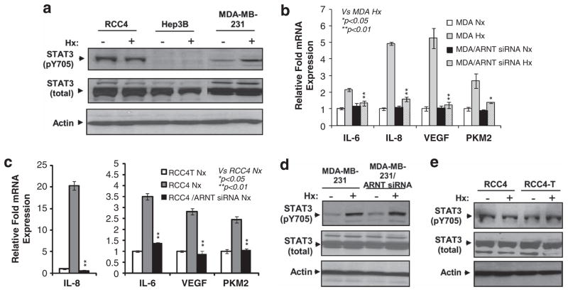
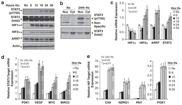
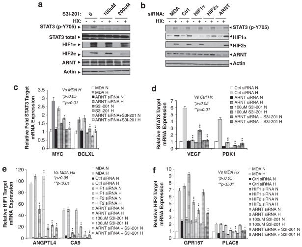
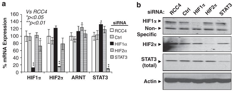
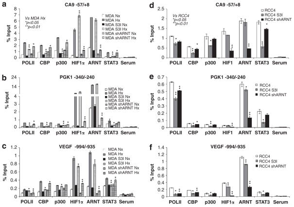
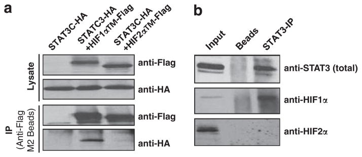
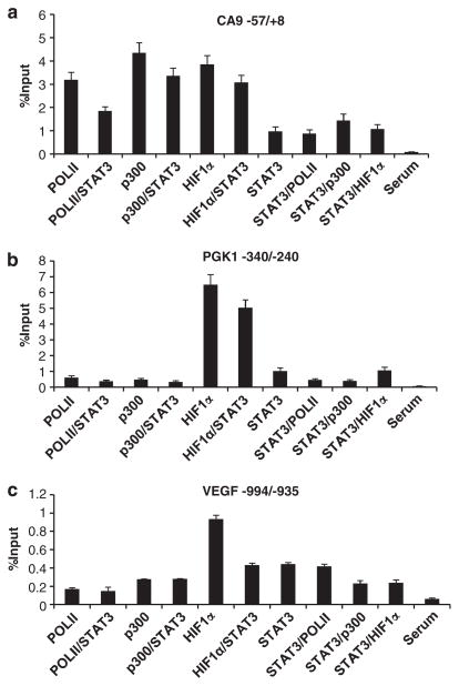
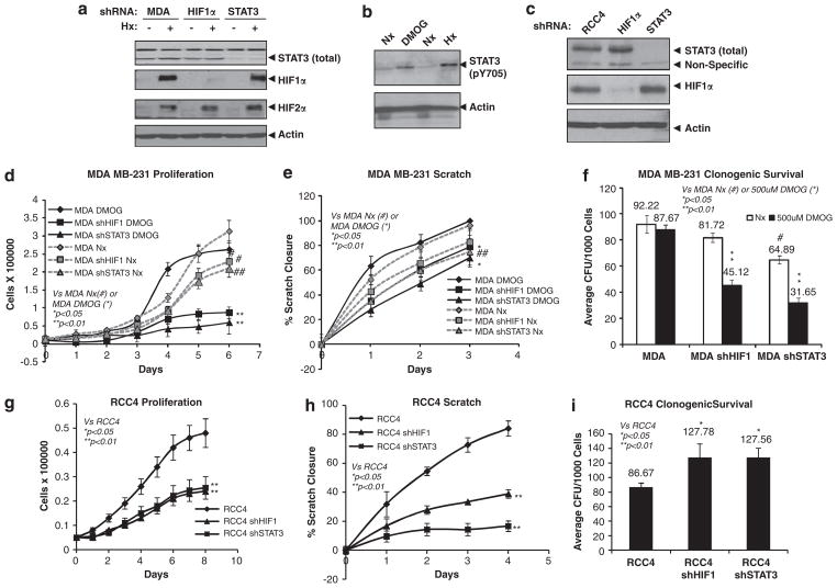
Solid tumors often exhibit simultaneously inflammatory and hypoxic microenvironments. The 'signal transducer and activator of transcription-3' (STAT3)-mediated inflammatory response and the hypoxia-inducible factor (HIF)-mediated hypoxia response have been independently shown to promote tumorigenesis through the activation of HIF or STAT3 target genes and to be indicative of a poor prognosis in a variety of tumors. We report here for the first time that STAT3 is involved in the HIF1, but not HIF2-mediated hypoxic transcriptional response. We show that inhibiting STAT3 activity in MDA-MB-231 and RCC4 cells by a STAT3 inhibitor or STAT3 small interfering RNA significantly reduces the levels of HIF1, but not HIF2 target genes in spite of normal levels of hypoxia-inducible transcription factor 1α (HIF1α) and HIF2α protein. Mechanistically, STAT3 activates HIF1 target genes by binding to HIF1 target gene promoters, interacting with HIF1α protein and recruiting coactivators CREB binding protein (CBP) and p300, and RNA polymerase II (Pol II) to form enhanceosome complexes that contain HIF1α, STAT3, CBP, p300 and RNA Pol II on HIF1 target gene promoters. Functionally, the effect of STAT3 knockdown on proliferation, motility and clonogenic survival of tumor cells in vitro is phenocopied by HIF1α knockdown in hypoxic cells, whereas STAT3 knockdown in normoxic cells also reduces cell proliferation, motility and clonogenic survival. This indicates that STAT3 works with HIF1 to activate HIF1 target genes and to drive HIF1-depedent tumorigenesis under hypoxic conditions, but also has HIF-independent activity in normoxic and hypoxic cells. Identifying the role of STAT3 in the hypoxia response provides further data supporting the effectiveness of STAT3 inhibitors in solid tumor treatment owing to their usefulness in inhibiting both the STAT3 and HIF1 pro-tumorigenic signaling pathways in some cancer types.


Conflict of interest statement

CONFLICT OF INTEREST

The authors declare no conflict of interest.

Figures

Figure 1
Figure 1
Hypoxia activates STAT3 activity in breast cancer cell line MDA-MB-231. (a) Western blot analysis of total and phosphorylated (Y705) STAT3 protein levels in normoxic and hypoxic (Hx) RCC4 (pVHL-null), Hep3B and MDA-MB-231 cancer cell lines. Beta-actin serves as loading control for this and other western blot analysis in the paper. (b) qPCR analysis of mRNA levels of reported STAT3 activators in normoxic and hypoxic MDA-MB-231 cells or cells targeted with ARNT siRNA. qPCR results were normalized to 18S rRNA expression and calibrated to normoxia control cells; error bars are ±1 s.d. from at least three independent experiments in this and other figures. Two-tail t-tests were performed for this and other studies in the paper with ‘*’ indicating P<0.05 and ‘**’ indicating P<0.01. Controls for the t-tests are indicated in the figures. (c) qPCR analysis of mRNA levels of reported STAT3 activators in normoxic RCC4 cells, RCC4 cells targeted with ARNT siRNA or normoxic pVHL-tranfected RCC4-T cells. (d) Western blot analysis of total and pY705STAT3 protein in normoxic and hypoxic MDA-MB-231 cells or MDA-MB-231 cells targeted with ARNT siRNA. (e) Western blot analysis of total and pY705STAT3 protein in normoxic or hypoxic RCC4 and RCC4 cells reconstituted with pVHL (RCC4-T cells).
Figure 2
Figure 2
Hypoxia-mediated STAT3 activation corresponds with maximal HIF target gene expression in MDA-MB-231 cells. (a) Western blot analysis of pY705STAT3, total STAT3, HIF1α, HIF2α and ARNT protein in MDA-MB-231 cells cultured under normoxia (Nx) or 6–30 h hypoxia (Hx). (b) Western blot analysis of pY705STAT3 and total STAT3 in nuclear (Nuc) and cytosolic (Cyt) fractions of the MDA-MB-231 cells cultured under Nx or 24 h Hx. (c) qPCR analysis of mRNA levels of HIF1α, HIF2α, ARNT and STAT3 in MDA-MB-231 cells cultured under Nx or 6–30 h of Hx. (d) qPCR analysis of mRNA levels of known STAT3/HIF common targets (PDK1 and VEGF) or STAT3 targets (MYC and BIRC5) in MDA-MB-231 cells under Nx and 6–30 h Hx. (e) qPCR analysis of mRNA levels of known HIF target genes in MDA-MB-231 cells under Nx and 6–30 h Hx.
Figure 3
Figure 3
Optimal hypoxic induction of HIF1 target genes in MDA-MB-231 cells requires STAT3 activity. (a) Western blot analysis of levels of pY705STAT3, total STAT3, HIF1α, HIF2α and ARNT protein in MDA-MB-231 cells cultured under normoxia (Nx) or 24 h hypoxia (Hx) in the absence or presence of 100 or 200 μM of the STAT3 inhibitor S3I-201. (b) Western blot analysis of indicated protein in Nx or 24 h Hx MDA-MB-231 cells (MDA), or MDA-MB-231 cells targeted with control siRNA or siRNA against HIF1α, HIF2α, or ARNT mRNA. (c) qPCR analysis of mRNA levels of known STAT3 target genes MYC and BCL-XL in Nx or 24 h Hx MDA cells, or cells targeted with ARNT siRNA, or with 100 μM STAT3 inhibitor S3I-201 or both ARNT siRNA and STAT3 inhibitor. (d) qPCR analysis of mRNA levels of known STAT3/HIF common target genes VEGF and PDK1 in Nx or 24 h Hx MDA cells as for Figures 3c, e and f) qPCR analysis of mRNA levels of (e) HIF1- and (f) HIF2-specific target genes in Nx or 24 h Hx MDA cells, siRNA-targeted MDA cells, 100 μM S3I-201-treated MDA cells or ARNT siRNA with STAT3 inhibitor.
Figure 4
Figure 4
HIF1α, HIF2α or STAT3 siRNA efficiently reduced mRNA and protein expression of HIF1α, HIF2α and STAT3 in RCC4 cells. (a) qPCR analysis of mRNA levels of HIF1α, HIF2α, ARNT and STAT3 in normoxic RCC4 cells and RCC4 cells targeted with HIF1α, HIF2α or STAT3 siRNA. (b) Western blot analysis of HIF1α, HIF2α or STAT3 protein levels in parental or siRNA-transfected RCC4 cells.
Figure 5
Figure 5
STAT3, but not HIF1α/ARNT is primarily responsible for recruiting CBP and p300 coactivators to the endogenous HIF1 target promoters. ChIP analysis of Pol II, CBP, p300, HIF1α, ARNT and STAT3 binding to the HIF1 target promoters around the reported HREs of the CA9 (a), PGK1 (b) and VEGF genes (c) in MDA-MB-231 cells under normoxic (Nx) or 24 h hypoxic (Hx) conditions or MDA cells treated with STAT3 inhibitor, or with ARNT stably knocked down. Co-precipitated DNA is presented as percent DNA compared with input controls and background level is quantified by DNA pulled down using non-reactive serum (Serum). Similar ChIP was performed in normoxic RCC4 cells, or RCC4 cells targeted with STAT3 inhibitor or ARNT stably knocked down for CA9 promoter (d), PGK1 promoter (e) and VEGF promoter (f).
Figure 6
Figure 6
STAT3 protein specifically interacts HIF1α, but not HIF2α protein. (a) Western blot analysis of Flag-tagged HIF1α and HIF2α proteins (anti-Flag) or HA-tagged STAT3 (anti-HA) in the lysates (Lysate) or anti-Flag M2 bead immunoprecipitated materials (IP) from 293T cells transfected with STAT3C-HA (HA-tagged constitutively active STAT3), STAT3C-HA +HIF1αTM-Flag (Flag tagged HIF1α with triple mutation to make HIF1α stable and active under normoxia), or STAT3C-HA +HIF2αTM-Flag. (b) Western blot analysis of STAT3, HIF1α or HIF2α protein in the RCC4 lysates (input) or IP materials from the protein-A/protein-G beads (beads) or IP materials using anti-STAT3 pY705 antibody and protein-A/protein-G beads.
Figure 7
Figure 7
STAT3, HIF1α, p300 and Pol II form a transcriptional complex on the endogenous HIF1 target promoters. ChIP/ReChIP analysis of STAT3, HIF1α, Pol II and P300 binding to the endogenous HIF1 target gene promoters CA9 (a), PGK1 (b) and VEGF (c) in 24 h hypoxic MDA-MB-231 cells. Antibodies used for the primary and secondary immunoprecipitation are listed as first/second. The co-precipitated DNA is presented in comparison with pull-down of each individual factor and non-reactive serum controls (Serum).
Figure 8
Figure 8
STAT3 knockdown phenocopies HIF1α knockdown in tumorigenic properties of MDA-MB-231 and RCC4 cells in vitro. (a) Western blot analysis of total STAT3, HIF1α and HIF2α in normoxic (Nx) or hypoxic (Hx) MDA-MB-231 cells or MDA-MB-231 cells with stably knocked down HIF1α or STAT3 protein. (b) Western blot analysis of pY705STAT3 in Nx or 24 h Hx MDA-MB-231 cells or Nx cells treated with or without 500 μM DMOG. (c) Western blot analysis of HIF1α or total STAT3 protein in RCC4 cells or RCC4 cells with HIF1α or STAT3 shRNA. (d) Proliferation of MDA-MB-231 cells, MDA/shHIF1α and MDA/shSTAT3 cells treated with or without 500 μM DMOG. (e) Motility of MDA-MB-231, MB-231/shHIF1 and MDA-MB-231/shSTAT3 cells grown with or without 500 μM DMOG was measured and presented as percent closure of a scratch assay. (f) Percent survival of colony-forming units of MDA-MB-231, MDA-MB-231/shHIF1 and MDA-MB-231/shSTAT3 cells grown in the presence or absence of 500 μM DMOG in a clonogenic survival assay. (g) Proliferation of RCC4, RCC4/shHIF1 and RCC4/shSTAT3 cells under Nx. (h) Motility of Nx RCC4, RCC4/shHIF1 and RCC4/shSTAT3 cells was measured and presented as percent closure in a scratch assay. (i) Percent survival of colony-forming units of RCC4, RCC4/shHIF1 and RCC4/shSTAT3 cells in a clonogenic survival assay.

Similar articles

Cited by

References

    1. Finger EC, Giaccia AJ. Hypoxia, inflammation, and the tumor microenvironment in metastatic disease. Cancer Metastasis Rev. 2010;29:285–293. - PMC - PubMed
    1. Keith B, Johnson RS, Simon MC. HIF1alpha and HIF2alpha: sibling rivalry in hypoxic tumour growth and progression. Nat Rev Cancer. 2012;12:9–22. - PMC - PubMed
    1. Semenza GL. Hypoxia-inducible factors in physiology and medicine. Cell. 2012;148:399–408. - PMC - PubMed
    1. Kaelin WG., Jr Cancer and altered metabolism: potential importance of hypoxia-inducible factor and 2-oxoglutarate-dependent dioxygenases. Cold Spring Harb Symp Quant Biol. 2011;76:335–345. - PMC - PubMed
    1. Bowman T, Garcia R, Turkson J, Jove R. STATs in oncogenesis. Oncogene. 2000;19:2474–2488. - PubMed

Publication types

MeSH terms

Substances