. 2013 May;31(5):458-64.
doi: 10.1038/nbt.2566. Epub 2013 Apr 21.

Identification of direct targets and modified bases of RNA cytosine methyltransferases

Affiliations

Identification of direct targets and modified bases of RNA cytosine methyltransferases

Vahid Khoddami et al. Nat Biotechnol. 2013 May.

Abstract

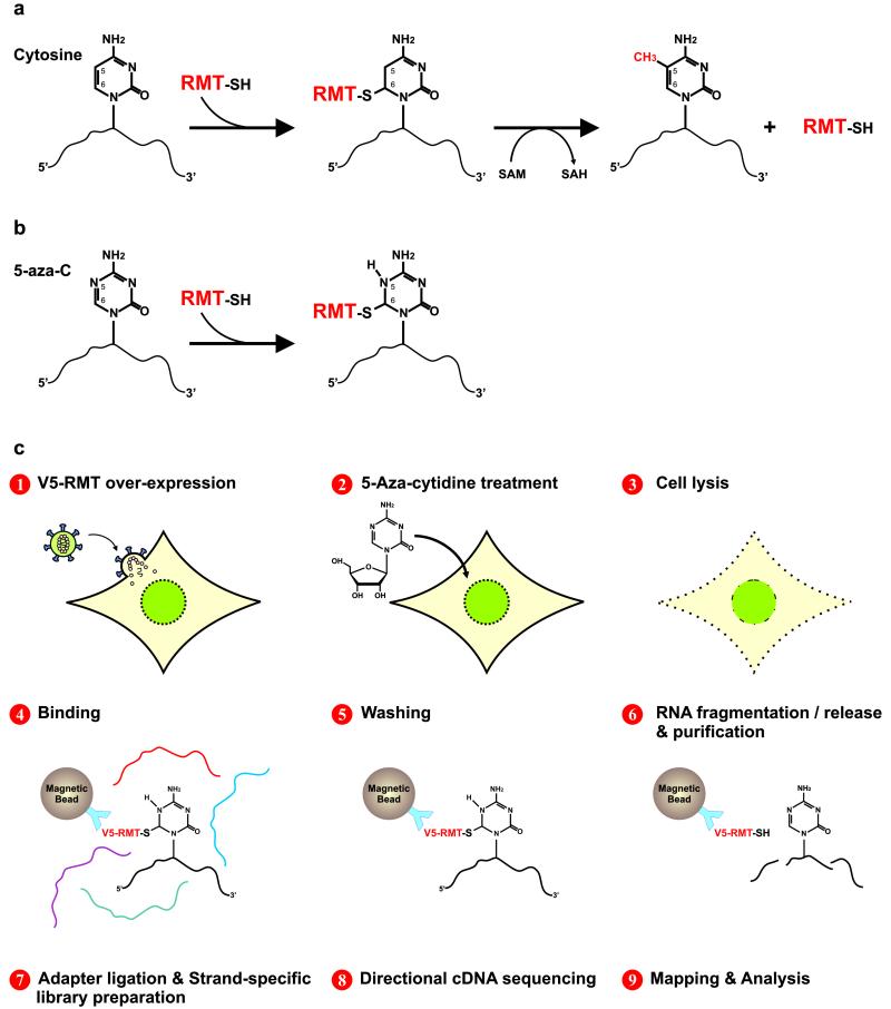
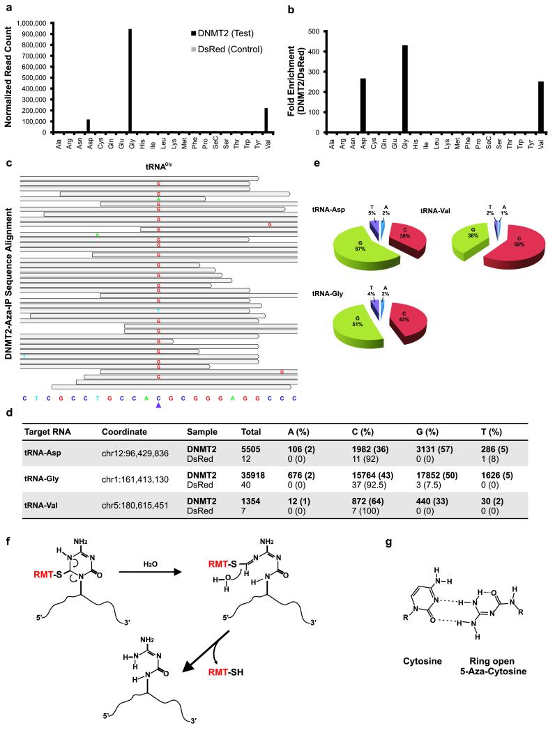
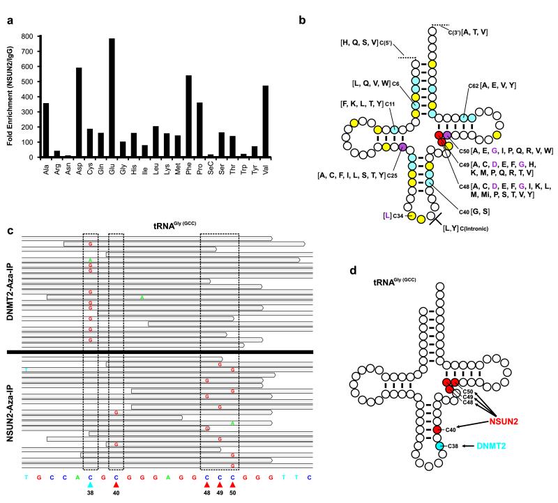
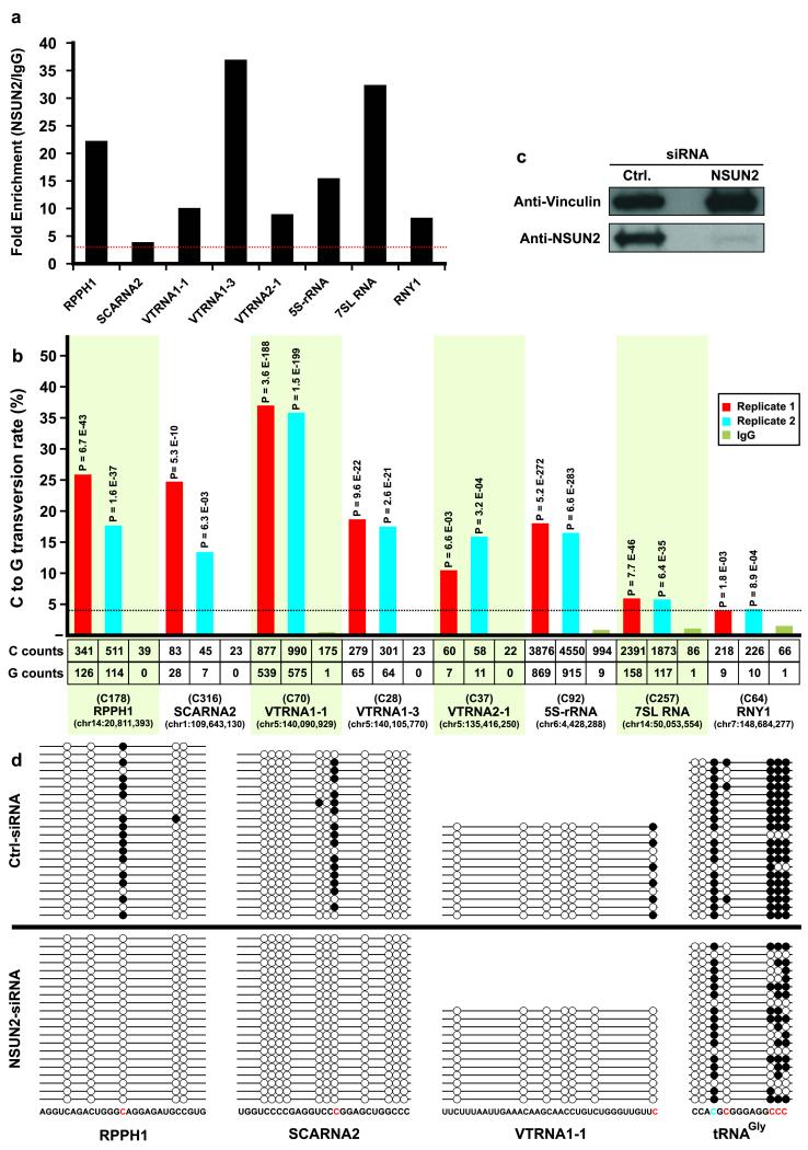
The extent and biological impact of RNA cytosine methylation are poorly understood, in part owing to limitations of current techniques for determining the targets of RNA methyltransferases. Here we describe 5-azacytidine-mediated RNA immunoprecipitation (Aza-IP), a technique that exploits the covalent bond formed between an RNA methyltransferase and the cytidine analog 5-azacytidine to recover RNA targets by immunoprecipitation. Targets are subsequently identified by high-throughput sequencing. When applied in a human cell line to the RNA methyltransferases DNMT2 and NSUN2, Aza-IP enabled >200-fold enrichment of tRNAs that are known targets of the enzymes. In addition, it revealed many tRNA and noncoding RNA targets not previously associated with NSUN2. Notably, we observed a high frequency of C→G transversions at the cytosine residues targeted by both enzymes, allowing identification of the specific methylated cytosine(s) in target RNAs. Given the mechanistic similarity of RNA cytosine methyltransferases, Aza-IP may be generally applicable for target identification.


Figures

Figure 1
Figure 1
RNA cytosine methylation mechanism and Aza-IP experimental design. (a) Schematic of m5C-RMTs catalyzing methylation of carbon 5 (C5) of cytosine. First, the enzyme forms a covalent thioester bond, connecting the cysteine residue of its catalytic domain to the C6 position of the target cytosine, forming an RMT-RNA adduct. Next, the RMT transfers a methyl group from cofactor S-Adenosyl Methionine (SAM) to the C5 of the target cytosine. The enzyme is then released from the adduct by β-elimination. Methylated RNA and S-Adenosyl-L-Homocysteine (SAH) are the product and byproduct of this reaction, respectively. (b) 5-azacytidine (5-aza-C) is a mechanism-based suicide inhibitor that traps the enzyme by forming a stable RMTRNA adduct. (c) Schematic representation of the Aza-IP technique.
Figure 2
Figure 2
Aza-IP analysis of DNMT2 RNA targets. (a) Graph depicts normalized reads mapping to each tRNA in the V5-DNMT2 (test) and V5-DsRed (control) datasets (one replicate of each shown). Each tRNA is designated by a three letter amino acid abbreviation. (b) Fold enrichment was calculated from the data shown in (a) by dividing the normalized RPKM (Reads Per Kilobase per Million mapped reads) values for each tRNA type in the V5-DNMT2 dataset by the values in the V5-DsRed dataset. (c) A representative snapshot from the Integrative Genomics Viewer (IGV, BROAD Inst.) browser depicting a subset of the sequencing reads mapped to a tRNAGly locus (chr1:161,413,119-161,413,141, human genome version 19 (hg19) bottom) at base pair resolution. The grey bars span the start/stop of individual sequencing reads mapped to the locus. The mismatched nucleotides are shown with colored letters and the matched nucleotides are hidden (grey). The purple arrowhead points to the tRNAGly C38 nucleotide (chr1:161,413,130). (d) Summary of the base distribution at the known DNMT2 target sites in tRNAAsp, tRNAGly and tRNAVal. The coordinate indicates the genomic location of the target cytosine in the human genome and the raw numbers are reported for both the V5-DNMT2 and V5-DsRed Aza-IP datasets. (e) Pie graphs showing the base distributions at the target nucleotide in the mapped reads. The numbers for the tRNAs are averaged over all of the annotated tRNA loci of same type in the human genome showing coverage over the target nucleotide (C38). (f) Schematic representation of the RMT-induced ring opening and RMT-RNA dissociation model. This model was proposed by Jackson-Grusby et al. for mammalian DNA cytosine methyltransferases and is adapted here for RMTs. RMT covalent linkage to the C6 position of 5-aza-C induces the rearrangement and ring opening and results in dissociation of the RMT from the target RNA molecule. (g) Base-pairing behavior of ring-open 5-aza-C. The ring-open 5-aza-C prefers to pair with cytosine and is therefore read as guanosine after RT-PCR and sequencing.
Figure 3
Figure 3
Aza-IP analysis of NSUN2 RNA targets. (a) Graph depicts fold enrichment of human tRNAs in V5-NSUN2 immunoprecipitates. Fold enrichment was calculated by dividing the normalized RPKM (Reads Per Kilobase per Million mapped reads) values for each tRNA type in the V5-NSUN2 replicate datasets (combined) by the values in the IgG control dataset. Each tRNA is designated by a three letter amino acid abbreviation. (b) A ‘standardized’ tRNA summarizing the human NSUN2 target cytosines revealed by Aza-IP in HeLa cells. Cytosines are color-coded based on the number of tRNA types that we found to be NSUN2 target sites: yellow, one tRNA type; blue, 2-5 tRNA types; purple, 6-9 tRNA types; and red >10 tRNA types. For each position, the individual tRNAs are designated by their single letter amino acid abbreviation, grouped in square brackets; purple letters refer to previously known NSUN2 target sites in the designated tRNA types (in human), , . For clarity of presentation, only selected positions are depicted (See Supplementary File 5 for all tRNAs, their isoacceptors/isodecoders, and target positions). (c) Integrative Genomics Viewer (IGV) browser snapshots of a random subset of the sequencing reads mapped to a tRNAGly(GCC) locus (chr17:8,029,095-8,029,117) from the separate DNMT2 (top) or NSUN2 Aza-IP (bottom) datasets. Stippled boxes show locations that meet target criteria with either enzyme. Arrowheads at bottom depict the sole DNMT2 target site (blue) or the four NSUN2 target sites (red). (d) A ‘standardized’ tRNAGly(GCC) with all five known m5C bases depicted, all five of which (and no other resident cytosine) were specifically and selectively identified by Aza-IP of DNMT2 or NSUN2.
Figure 4
Figure 4
New ncRNA targets and sites for human NSUN2, and validation through siRNA knockdown and bisulfite sequencing. (a) Graph depicts fold enrichment of human ncRNAs in V5-NSUN2 immunoprecipitates. Fold enrichment was calculated by dividing the normalized RPKM (Reads Per Kilobase per Million mapped reads) values for each ncRNA in the V5-NSUN2 replicate datasets (combined) by the values in the IgG control dataset. The red horizontal dotted line shows the 3-fold enrichment cut-off criteria. (b) C>G transversion at particular sites within ncRNAs enriched in anti-V5-NSUN2 immunoprecipitates. For each of the eight enriched ncRNAs, we depict the single target site with the highest C>G transversion (statistics for other candidate sites in Supplementary File 5). The bar graph shows the C>G transversion occurrence (% of total) in each V5-NSUN2 Aza-IP replicate and IgG only control; the horizontal dotted line shows the 4% transversion cut-off. P-values calculated by VarScan are indicated above columns. Provided underneath are the total number of sequenced C and G nucleotides (counts), the base position of the target cytosine within the ncRNA, and its encoded location in the genome (hg19). (c) Extracts (60μg of protein was loaded on the gel) of HeLa cells expressing NSUN2 or non-specific (control) siRNA pools were probed with anti-hNSUN2 or anti-Vinculin (control). (d) Total RNA was extracted from HeLa cells treated with NSUN2 or control siRNAs; RNA was subjected to bisulfite treatment, PCR amplification, cloning and sequencing of several clones per sample (see Online Methods). Candidate target sites of NSUN2 (in red; C178 in RPPH1, C316 in SCARNA2, C70 in VTRNA1-1, and C40, C48, C49 and C50 in tRNAGly(GCC)) and DNMT2 (in blue; C38 in tRNAGly(GCC)) were analyzed.

Similar articles

Cited by

References

    1. Motorin Y, Lyko F, Helm M. 5-methylcytosine in RNA: detection, enzymatic formation and biological functions. Nucleic Acids Res. 2010;38:1415–1430. - PMC - PubMed
    1. Squires JE, et al. Widespread occurrence of 5-methylcytosine in human coding and non-coding RNA. Nucleic Acids Res. 2012 - PMC - PubMed
    1. Jurkowski TP, et al. Human DNMT2 methylates tRNA(Asp) molecules using a DNA methyltransferase-like catalytic mechanism. RNA. 2008;14:1663–1670. - PMC - PubMed
    1. King MY, Redman KL. RNA methyltransferases utilize two cysteine residues in the formation of 5-methylcytosine. Biochemistry. 2002;41:11218–11225. - PubMed
    1. Goll MG, et al. Methylation of tRNAAsp by the DNA methyltransferase homolog Dnmt2. Science. 2006;311:395–398. - PubMed

Publication types

Associated data