. 2013 May;31(5):440-7.
doi: 10.1038/nbt.2565. Epub 2013 Apr 21.

Medial ganglionic eminence-like cells derived from human embryonic stem cells correct learning and memory deficits

Affiliations

Medial ganglionic eminence-like cells derived from human embryonic stem cells correct learning and memory deficits

Yan Liu et al. Nat Biotechnol. 2013 May.

Abstract

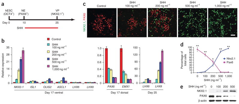
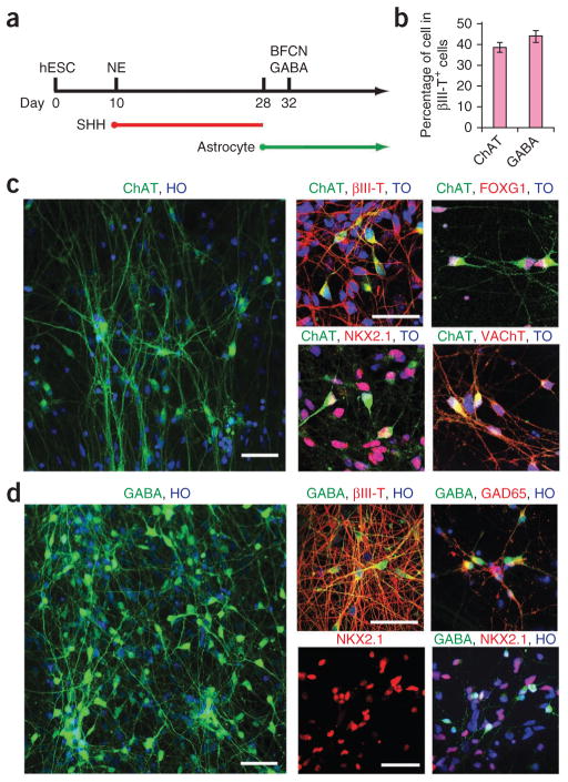
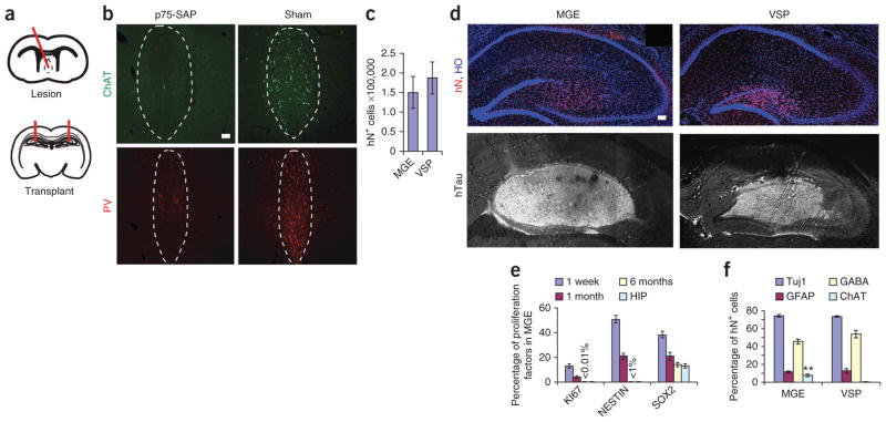
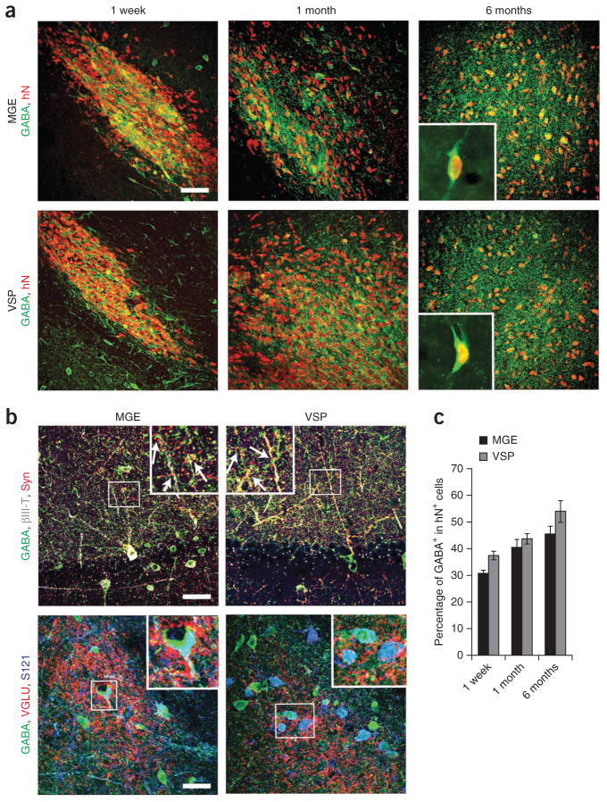
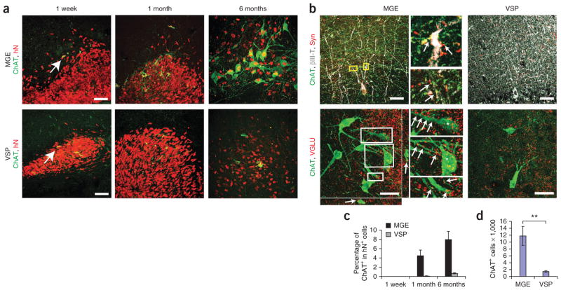
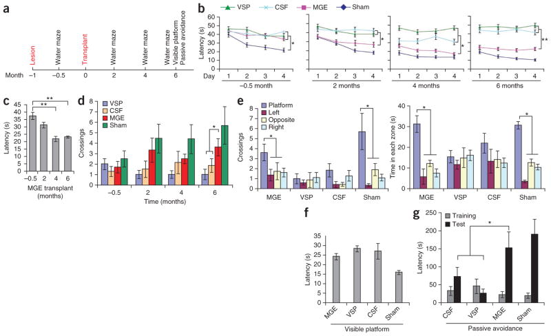
Dysfunction of basal forebrain cholinergic neurons (BFCNs) and γ-aminobutyric acid (GABA) interneurons, derived from medial ganglionic eminence (MGE), is implicated in disorders of learning and memory. Here we present a method for differentiating human embryonic stem cells (hESCs) to a nearly uniform population of NKX2.1(+) MGE-like progenitor cells. After transplantation into the hippocampus of mice in which BFCNs and some GABA neurons in the medial septum had been destroyed by mu P75-saporin, human MGE-like progenitors, but not ventral spinal progenitors, produced BFCNs that synaptically connected with endogenous neurons, whereas both progenitors generated similar populations of GABA neurons. Mice transplanted with MGE-like but not spinal progenitors showed improvements in learning and memory deficits. These results suggest that progeny of the MGE-like progenitors, particularly BFCNs, contributed to learning and memory. Our findings support the prospect of using human stem cell-derived MGE-like progenitors in developing therapies for neurological disorders of learning and memory.


Conflict of interest statement

COMPETING FINANCIAL INTERESTS

The authors declare no competing financial interests.

Figures

Figure 1
Figure 1
SHH-dependent specification of basal forebrain progenitors from hESCs. (a) Schematic showing specification of NKX2.1+ ventral progenitors (VP) from neuroepithelia (NE) by SHH for 2 weeks starting from day 10. (b) Quantitative RT-PCR analysis of various markers (expression relative to control: 0 ng ml−1 SHH treatment group) on indicated days of differentiation. Dorsal and ventral refer to dorsal and ventral forebrain markers. Error bars, s.e.m. (c) Immunocytochemistry analysis of hESC-derived neural progenitors for NKX2.1 and PAX6 expression in response to SHH on day 25 of differentiation. Scale bar, 50 μm. (d) Quantification of data in c. Error bars, s.e.m.; *P < 0.05 and **P < 0.01. (e) Western blot analysis of hESC-derived neural progenitor lysates with antibodies to indicated proteins in response to SHH treatment on day 25 after differentiation.
Figure 2
Figure 2
Differentiation of basal forebrain progenitors to cholinergic neurons and GABAergic interneurons. (a) Schematic of neuronal differentiation from basal forebrain progenitors. NE, neuroepithelia. (b) Quantification of the percentage of ChAT+ and GABA+ neurons in βIII-tubulin+ (βIII-T+) cells. Error bars, s.e.m. (c) Immunofluorescence images of ChAT+ cholinergic neurons immunostained for βIII-tubulin (βIII-T), VAChT, NKX2.1 and FOXG1. (d) Immunofluorescence images of GABA neurons immunostained for βIII-tubulin (βIII-T), NKX2.1 and GAD65. TO, topo3; HO, Hoechst staining. Scale bars, 50 μm.
Figure 3
Figure 3
Survival, proliferation and differentiation of grafted cells in lesioned hippocampi. (a) Schematics showing medial septal lesion and hippocampal transplantation sites. (b) Immunofluorescence images showing the expression of ChAT and parvalbumin (PV) in the p75-SAP–injected medial septum and the CSF injection control (sham). Dashed outlines show medial septum area. (c) Total grafted cell numbers (human nuclear protein–immunopositive; hN+) in MGE and VSP groups. (d) Immunofluorescence images of human cells showing hN and human neural fibers (hTau) in MGE and VSP grafts. Hoechst (HO)-stained cell nuclei reveal hippocampal layer structures. Scale bars, 50 μm (b, d). (e) Proportion of KI67+, NESTIN+ and SOX2+ cells among total human (hN+) cells 1 week, 1 month and 6 months after transplantation in MGE as well as Sox2+ cells in adult SCID mouse hippocampus (HIP). Values for KI67 and NESTIN at 6 months are too small to be shown with bars and are labeled with values. (f) Quantification of neural subtypes among total grafted human (hN+) cells by 6 months. **P < 0.01. Error bars, s.e.m.
Figure 4
Figure 4
Differentiation and integration of grafted GABA+ neurons. (a) Immunofluorescence images of GABA+ neurons among human (human nuclear protein–immunopositive; hN+) cells over 1 week, 1 month and 6 months after transplantation. Insets, higher magnification of GABA+ and human nuclear protein–immunopositive (hN+) neurons in grafts 6 months after transplantation. (b) Immunostaining images showing GABA, human synaptophysin (Syn) and βIII-tubulin (βIII-T) (top) and VGLU punctae, GABA and STEM121 (S121) (bottom) for MGE and VSP groups. Insets, magnified views of boxed regions (arrows indicate colocalization). Scale bars, 50 μm. (c) Quantification of GABAergic neurons among total grafted cells in the MGE and VSP groups at indicated times. Error bars, s.e.m.
Figure 5
Figure 5
Differentiation and integration of cholinergic neurons. (a) Immunostaining of ChAT+ neurons over time in MGE and VSP groups. hN, human nuclear protein. (b) Immunostaining images of ChAT, human synaptophysin (Syn) and βIII-tubulin (βIII-T) (top) and of ChAT and VGLU (bottom) for MGE and VSP groups. Insets, magnified views of boxed regions (arrows indicate colocalization). Scale bars, 50 μm. (c) Percentage of human ChAT+ neurons among total grafted human cells 1 week, 1 month and 6 months after transplantation. hN, human nuclear protein. (d) Quantification of total ChAT+ cell numbers in MGE and VSP groups. Error bars, s.e.m; **P < 0.01.
Figure 6
Figure 6
Transplantation of hESC-derived MGE progenitors contributes to functional recovery. (a) Timeline of surgery and behavioral analyses. (b) Morris water-maze test–based analysis of latency in finding a hidden platform before transplantation in mice treated as indicated. Sham: aCSF injection in medial septum. After transplantation, the MGE group, but not the VSP or CSF group, progressively shortened the latency in finding the platform: −0.5 months, F = 19.434 and P = 0.0006; 2 months, F = 5.671 and P = 0.01; 4 months, F = 12.14 and P = 0.0002; 6 months, F = 9.966 and P = 0.0007. (c) Summary of the latency in finding the hidden platform for the MGE group over time. (d) Times crossing the removed platform in the MGE-transplanted group compared to controls 6 months after transplantation. F = 3.359, P = 0.048. (e) Differential crossing and time spent in each of the quadrants by the mice. MGE crossings, F = 3.034 and P = 0.046; time, MGE time spent in quadrant, F = 12.093 and P < 0.001. (f) Latency in landing on the visible platform. (g) Latency in passive avoidance tests. F = 5.257 and P = 0.0159. Error bars, s.e.m.; *P < 0.05, **P < 0.01.

Similar articles

Cited by

References

    1. Sussel L, Marin O, Kimura S, Rubenstein JL. Loss of Nkx2.1 homeobox gene function results in a ventral to dorsal molecular respecification within the basal telencephalon: evidence for a transformation of the pallidum into the striatum. Development. 1999;126:3359–3370. - PubMed
    1. Rubenstein JL, Shimamura K, Martinez S, Puelles L. Regionalization of the prosencephalic neural plate. Annu Rev Neurosci. 1998;21:445–477. - PubMed
    1. Wilson SW, Rubenstein JL. Induction and dorsoventral patterning of the telencephalon. Neuron. 2000;28:641–651. - PubMed
    1. Zhao Y, et al. The LIM-homeobox gene Lhx8 is required for the development of many cholinergic neurons in the mouse forebrain. Proc Natl Acad Sci USA. 2003;100:9005–9010. - PMC - PubMed
    1. Campbell K. Dorsal-ventral patterning in the mammalian telencephalon. Curr Opin Neurobiol. 2003;13:50–56. - PubMed

Publication types