

Molecular components of the Mammalian circadian clock
- PMID: 23604473
- PMCID: PMC3762864
- DOI: 10.1007/978-3-642-25950-0_1
Molecular components of the Mammalian circadian clock
Abstract
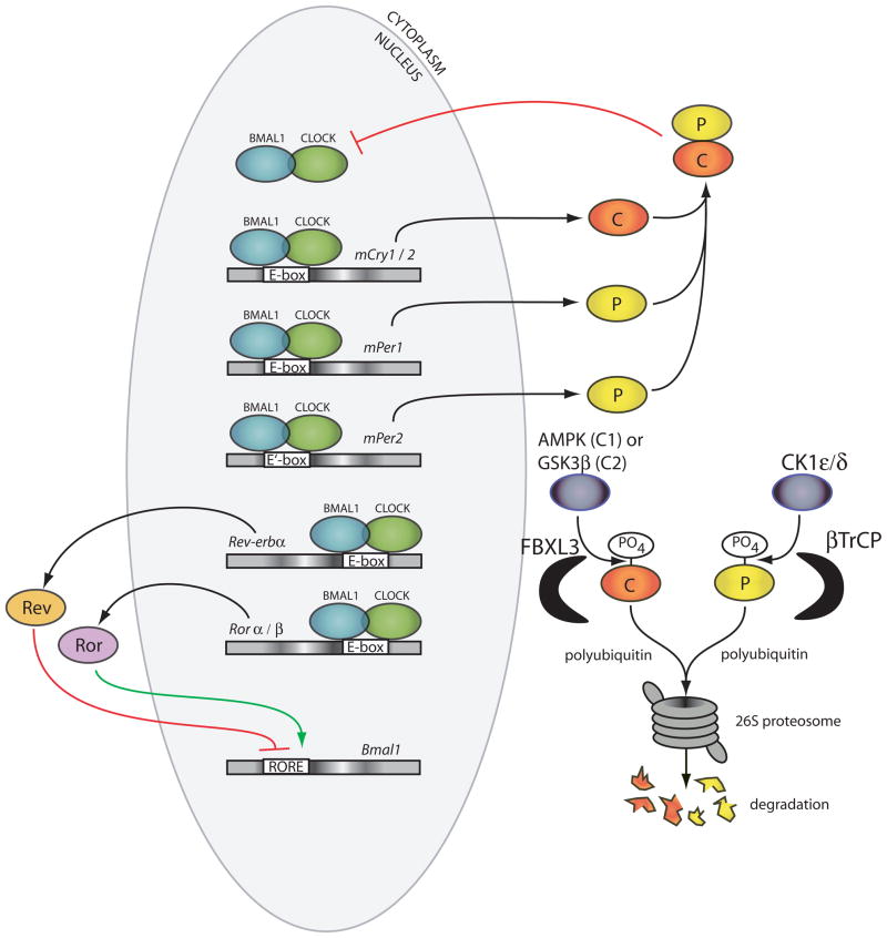
Mammals synchronize their circadian activity primarily to the cycles of light and darkness in the environment. This is achieved by ocular photoreception relaying signals to the suprachiasmatic nucleus (SCN) in the hypothalamus. Signals from the SCN cause the synchronization of independent circadian clocks throughout the body to appropriate phases. Signals that can entrain these peripheral clocks include humoral signals, metabolic factors, and body temperature. At the level of individual tissues, thousands of genes are brought to unique phases through the actions of a local transcription/translation-based feedback oscillator and systemic cues. In this molecular clock, the proteins CLOCK and BMAL1 cause the transcription of genes which ultimately feedback and inhibit CLOCK and BMAL1 transcriptional activity. Finally, there are also other molecular circadian oscillators which can act independently of the transcription-based clock in all species which have been tested.
Figures
References
-
- Aschoff J. Circadian control of body temperature. Journal of Thermal Biology. 1983;8:143–147.
-
- Cameron MA, Barnard AR, Lucas RJ. The electroretinogram as a method for studying circadian rhythms in the mammalian retina. J Genet. 2008;87:459–466. - PubMed
-
- Reid KJ, Zee PC. Circadian rhythm disorders. Semin Neurol. 2009;29:393–405. - PubMed
Publication types
MeSH terms
Substances
Grants and funding
LinkOut - more resources
Full Text Sources
Other Literature Sources
Medical
Molecular Biology Databases
