

Occurrence of gustducin-immunoreactive cells in von Ebner's glands of guinea pigs
- PMID: 23604549
- PMCID: PMC3824220
- DOI: 10.1007/s00418-013-1094-9
Occurrence of gustducin-immunoreactive cells in von Ebner's glands of guinea pigs
Abstract
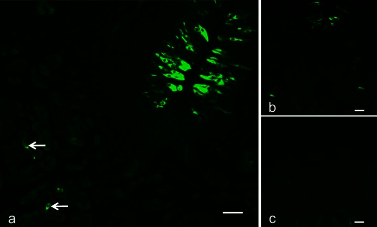
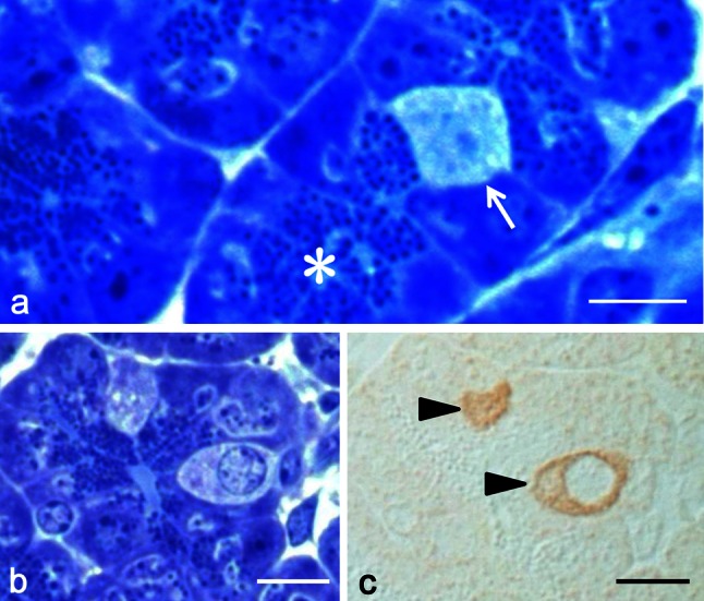
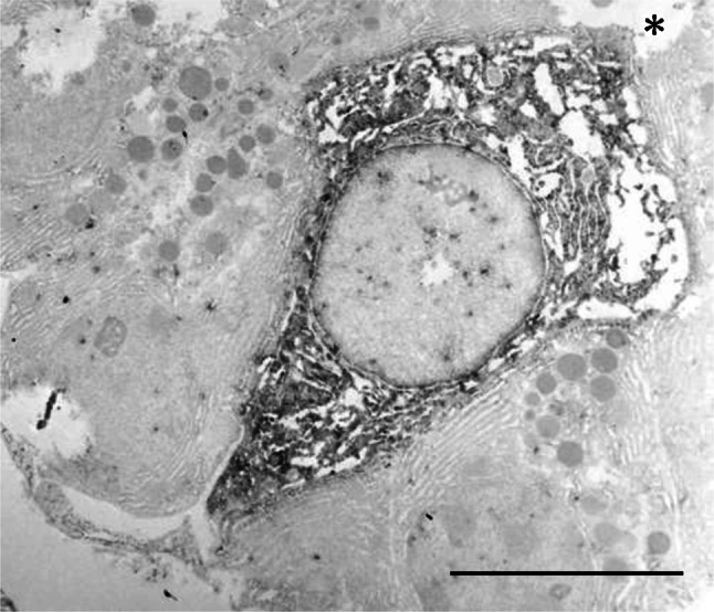
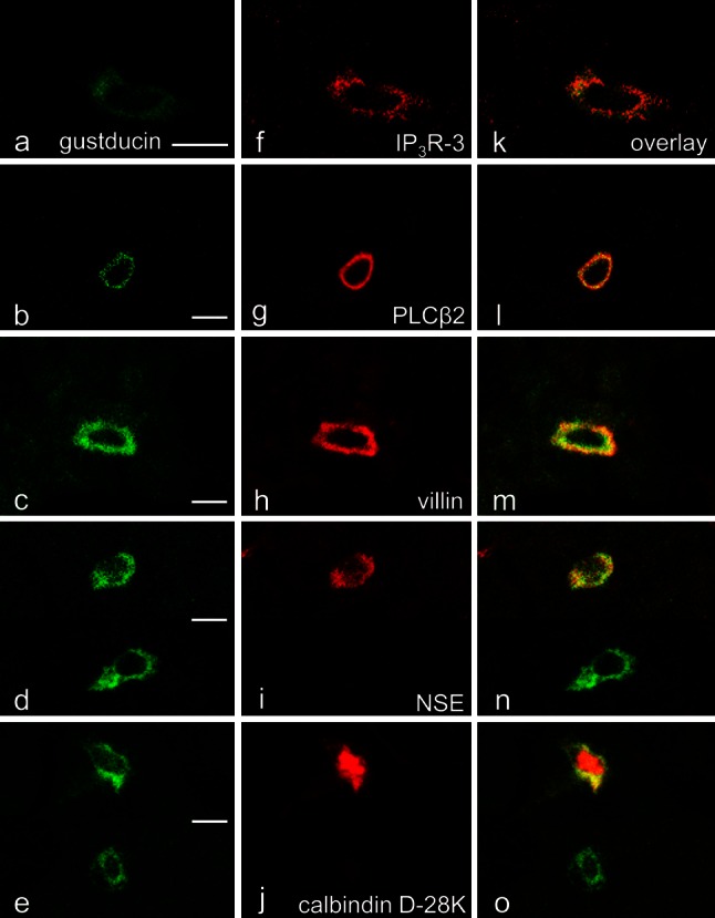
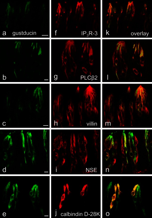
An immunohistochemical examination of guinea-pig taste buds in vallate papillae revealed gustducin-immunoreactive cells in the area of von Ebner's glands, minor salivary glands. Since there have been no reports describing those cells in these locations for other species, we investigated these glands in order both to localize the cells and compare their immunoreactive characteristics with corresponding cells in the vallate taste buds. The gustducin-immunoreactive cells coincided with cells containing no secretory granules in the end portion of the glands, which was supported by the electron-microscopic immunocytochemistry. Double immunofluorescence microscopy confirmed these cells to be entirely immunopositive to type III inositol 1,4,5-triphosphate receptor (IP3R-3), phospholipase Cβ2 (PLCβ2), and villin and also partly immunopositive to neuron-specific enolase (NSE) and calbindin D-28K. The gustducin-immunoreactive cells in the vallate taste buds exhibited completely the same immunoreactivities for these five molecules. Accordingly, the present results give credence to a consideration that the gustducin-immunnoreactive cells in both locations are identical in function(s) e.g., chemo-reception.
Figures
Similar articles
-
Existence of subtypes of gustducin-immunoreactive cells in the vallate taste bud of guinea pigs.Arch Histol Cytol. 2007 Dec;70(5):291-6. doi: 10.1679/aohc.70.291. Arch Histol Cytol. 2007. PMID: 18431029
-
Alpha-gustducin-immunoreactive solitary chemosensory cells in the developing chemoreceptorial epithelium of the rat vallate papilla.Chem Senses. 1999 Oct;24(5):469-72. doi: 10.1093/chemse/24.5.469. Chem Senses. 1999. PMID: 10576254
-
Human taste cells express the G protein alpha-gustducin and neuron-specific enolase.Brain Res Mol Brain Res. 1994 Mar;22(1-4):193-203. doi: 10.1016/0169-328x(94)90047-7. Brain Res Mol Brain Res. 1994. PMID: 8015379
-
Differentiation of the lingual and palatal gustatory epithelium of the rat as revealed by immunohistochemistry of alpha-gustducin.Arch Histol Cytol. 2001 Oct;64(4):401-9. doi: 10.1679/aohc.64.401. Arch Histol Cytol. 2001. PMID: 11757909
-
Fixation conditions affect the immunoreactivity of gustducin in rat vallate taste buds.Arch Histol Cytol. 2010;73(2):91-3. doi: 10.1679/aohc.73.91. Arch Histol Cytol. 2010. PMID: 21566335
Cited by
-
The Histochem Cell Biol conspectus: the year 2013 in review.Histochem Cell Biol. 2014 Apr;141(4):337-63. doi: 10.1007/s00418-014-1207-0. Epub 2014 Mar 9. Histochem Cell Biol. 2014. PMID: 24610091 Free PMC article. Review.
References
-
- Azzali G, Gatti R, Bucci G, Orlandini G. Fine structure of bat deep posterior lingual glands (von Ebner’s) J Submicrosc Cytol Pathol. 1989;21:669–684. - PubMed
Publication types
MeSH terms
Substances
LinkOut - more resources
Full Text Sources
Other Literature Sources
