. 2013 Jul;48(1):85-92.
doi: 10.1002/mus.23699. Epub 2013 Apr 21.

Spasticity, weakness, force variability, and sustained spontaneous motor unit discharges of resting spastic-paretic biceps brachii muscles in chronic stroke

Affiliations

Spasticity, weakness, force variability, and sustained spontaneous motor unit discharges of resting spastic-paretic biceps brachii muscles in chronic stroke

Shuo-Hsiu Chang et al. Muscle Nerve. 2013 Jul.

Abstract

Introduction: The purpose of our study was to examine relations among spasticity, weakness, force variability, and sustained spontaneous motor unit discharges in spastic-paretic biceps brachii muscles in chronic stroke.

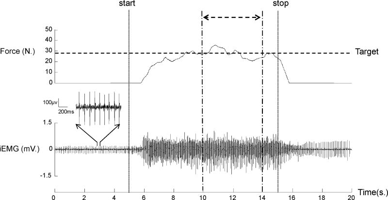
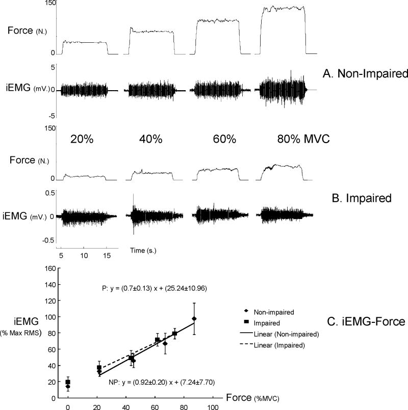
Methods: Ten chronic stroke subjects produced submaximal isometric elbow flexion force on impaired and non-impaired sides. Intramuscular EMG (iEMG) was recorded from biceps and triceps brachii muscles.

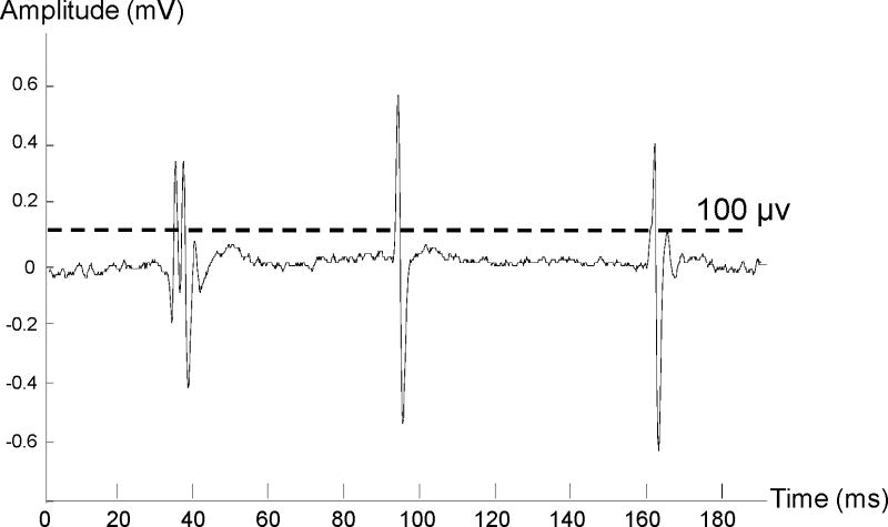
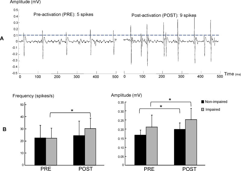
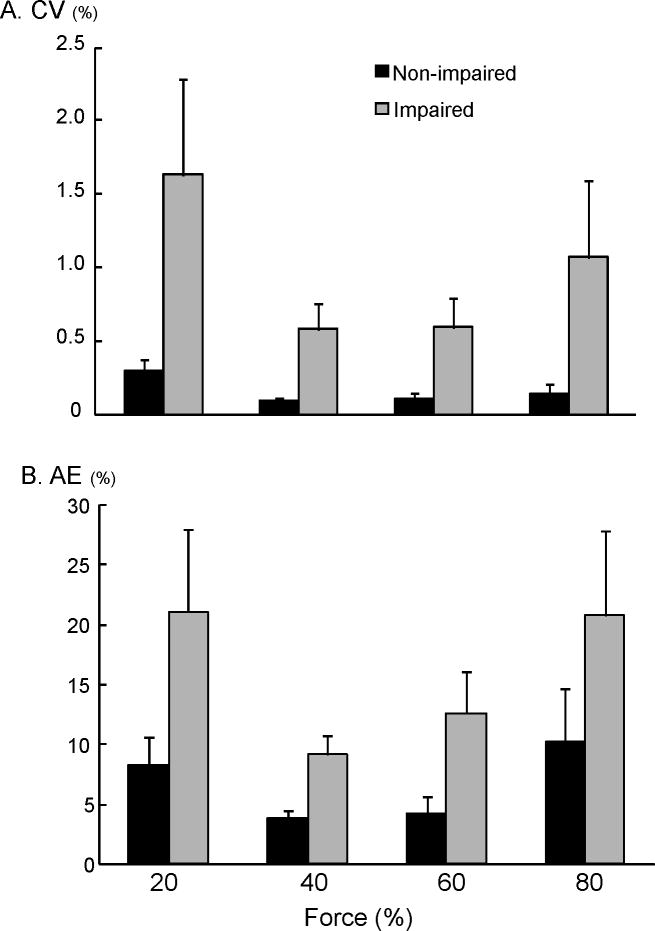
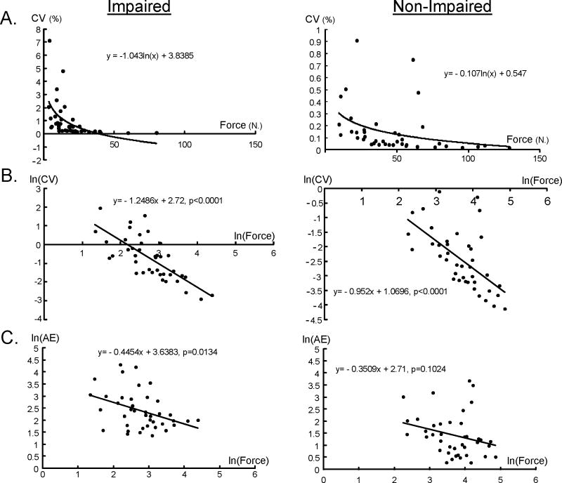
Results: We observed sustained spontaneous motor unit discharges in resting biceps on iEMG. Spontaneous discharges increased after voluntary activation only on the impaired side. The impaired side had greater matching errors and greater fluctuations in isometric force. Spontaneous discharges were not related functionally to spasticity, force variability, or weakness. However, greater strength on the impaired side correlated with less force variability.

Conclusion: Weakness rather than spasticity is a main factor interfering with voluntary force control in paretic-spastic biceps brachii muscles in chronic stroke.


Figures

Figure 1
Figure 1
A sample trace of spontaneous motor unit discharges. A threshold of 100 μV was used to detected spontaneous motor unit spikes.
Figure 2
Figure 2
A representative trial. The subject performed an isometric elbow flexion after the start signals (vertical dash line) to generate a submaximal force to match the pre-set target level (80% MVC, horizontal dotted line) as accurately as possible for 10s. The subject was asked to relax the muscle after the stop signal (vertical dotted line). In this trial, sustained spontaneous motor unit discharges were observed both before and after activation. Variables of EMG and force control were calculated in the 4-s period (double head arrow) during the steady force generation. The magnified motor unit activities in 1-s baseline (3rd – 4ths) are displayed. iEMG: intramuscular EMG.
Figure 3
Figure 3
Effect of voluntary contraction on spontaneous motor unit discharges. The number of spikes and amplitudes of spontaneous discharges increased on the impaired side in a typical trial (panel A). The averaged results (panel B) showed increased frequency and amplitude of spontaneous discharges after voluntary contraction on the impaired side, while only amplitude increased after voluntary contraction on the non-impaired side. Asterisk indicates p<0.05.
Figure 4
Figure 4
Representative raw EMG and force signals during submaximal force production from a typical subject (A and B). Raw EMG amplitudes were higher in the non-impaired side in comparison to the impaired side Raw EMGs increased with increasing force in both biceps. Average normalized EMG-force relations (C) The normalized iEMG amplitudes at rest (no force generated) were shown but not included in estimation of EMG-force relations. iEMG: intramuscular EMG.
Figure 5
Figure 5
Impaired voluntary force control. The spastic-paretic biceps has greater fluctuation and greater matching errors as compared to the non-impaired side across all force levels. NP: non-impaired; P: impaired.
Figure 6
Figure 6
The relationship between force and CV and AE in non-impaired and impaired limbs. The data are plotted on a linear axis in (A), and the natural log of the same data is plotted in (B). The natural log of the force and AE is plotted in (C).

References

    1. Lukacs M. Electrophysiological signs of changes in motor units after ischaemic stroke. Clin Neurophysiol. 2005;116:1566–1570. - PubMed
    1. Lewek MD, Hornby TG, Dhaher YY, Schmit BD. Prolonged quadriceps activity following imposed hip extension: a neurophysiological mechanism for stiff-knee gait? J Neurophysiol. 2007;98:3153–3162. - PMC - PubMed
    1. Mottram CJ, Suresh NL, Heckman CJ, Gorassini MA, Rymer WZ. Origins of abnormal excitability in biceps brachii motoneurons of spastic-paretic stroke survivors. J Neurophysiol. 2009;102:2026–2038. - PMC - PubMed
    1. Mottram CJ, Wallace CL, Chikando CN, Rymer WZ. Origins of spontaneous firing of motor units in the spastic-paretic biceps brachii muscle of stroke survivors. J Neurophysiol. 2010;104:3168–3179. - PMC - PubMed
    1. Seo NJ, Rymer WZ, Kamper DG. Delays in grip initiation and termination in persons with stroke: effects of arm support and active muscle stretch exercise. J Neurophysiol. 2009;101:3108–3115. - PubMed

Publication types

MeSH terms