. 2013 Jul;65(7):1843-52.
doi: 10.1002/art.37977.

Glucosamine activates autophagy in vitro and in vivo

Affiliations

Glucosamine activates autophagy in vitro and in vivo

Beatriz Caramés et al. Arthritis Rheum. 2013 Jul.

Abstract

Objective: Aging-associated changes in articular cartilage represent a main risk factor for osteoarthritis (OA). Autophagy is an essential cellular homeostasis mechanism. Aging-associated or experimentally induced defects in autophagy contribute to organismal- and tissue-specific aging, while enhancement of autophagy may protect against certain aging-related pathologies such as OA. The objective of this study was to determine whether glucosamine can activate autophagy.

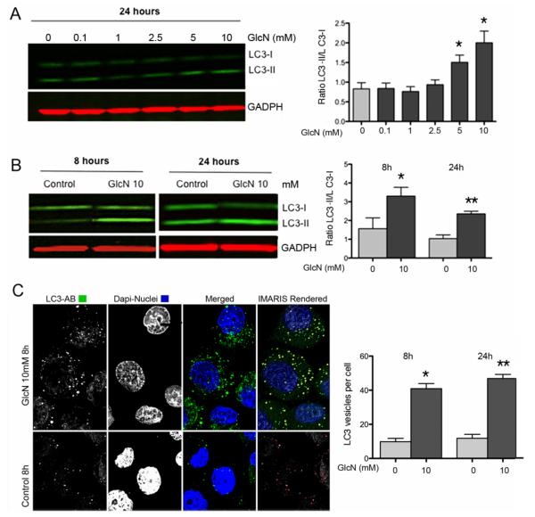
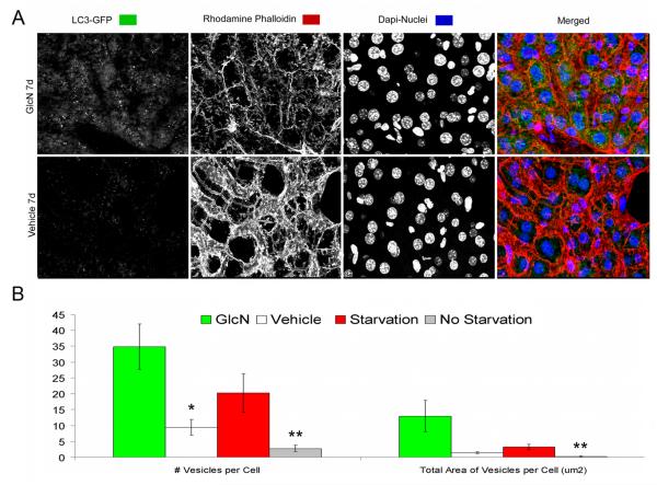
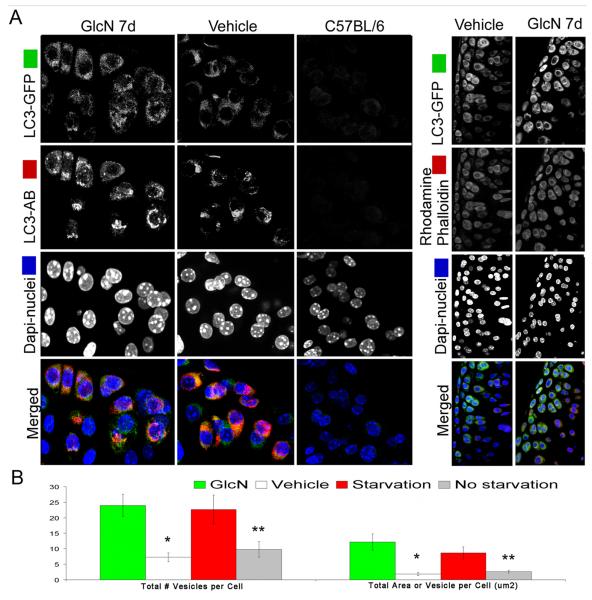
Methods: Chondrocytes from normal human articular cartilage were treated with glucosamine (0.1- 10 mM). Autophagy activation and phosphorylation levels of Akt, FoxO3, and ribosomal protein S6 were determined by Western blotting. Autophagosome formation was analyzed by confocal microscopy. Reporter mice systemically expressing green fluorescent protein (GFP) fused to light chain 3 (LC3) (GFP-LC3-transgenic mice) were used to assess changes in autophagy in response to starvation and glucosamine treatment.

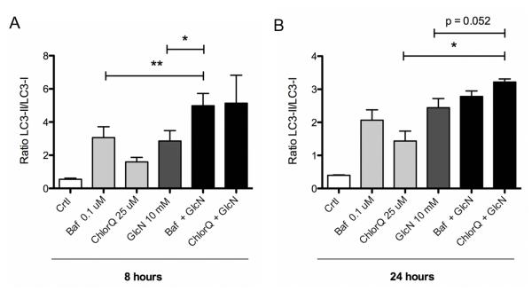
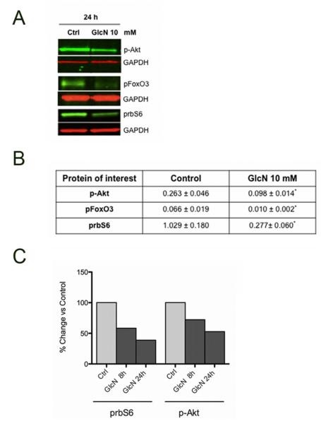
Results: Glucosamine treatment of chondrocytes activated autophagy, as indicated by increased LC3-II levels, formation of LC3 puncta, and increased LC3 turnover. This was associated with glucosamine-mediated inhibition of the Akt/FoxO3/mammalian target of rapamycin pathway. Administration of glucosamine to GFP-LC3-transgenic mice markedly activated autophagy in articular cartilage.

Conclusion: Glucosamine modulates molecular targets of the autophagy pathway in vitro and in vivo, and the enhancement of autophagy is mainly dependent on the Akt/FoxO/mTOR pathway. These findings suggest that glucosamine is an effective autophagy activator and should motivate future studies on the efficacy of glucosamine in modifying aging-related cellular changes and supporting joint health.


Figures

Figure 1
Figure 1. GlcN and autophagy activation in human articular chondrocytes
A, Normal human chondrocytes were untreated (DMEM 2% CS) or treated with GlcN (0.1, 1, 2.5, 5 and 10 mM) for 24 h. Total protein was analyzed by western blotting using anti-LC3 and GAPDH. Values represent mean ± SD of three separate experiments, * = P < 0.01 vs. control. Bar graphs show quantification of western signals expressed as LC3II/LC3I ratios. B, Normal human chondrocytes were untreated (DMEM 2% CS) or treated with GlcN (10 mM) for 8 or 24 h. LC3 was analyzed by western blotting. C, Normal human chondrocytes were untreated (DMEM 2% CS) or treated with GlcN (10 mM) for 8 and 24 hours stained with anti-LC3 and analyzed by confocal microscopy. The right-upper panel series represents a 3D IMARIS rendered reconstruction of the left-panels. The right-bottom panel represents vesicles that were outlined by Image Pro software (outlined in red) for quantification of vesicles. Magnification: 63x. The bar graph shows quantification of LC3-positive vesicles at 8 and 24 hours.
Figure 2
Figure 2. GlcN increases autophagy flux in human articular chondrocytes
A, Normal human chondrocytes were untreated (DMEM 2% CS) or treated with 10 mM GlcN for 8h or 24h, with or without Bafilomycin (Baf; 0.1 μM) or Chloroquine (ChlorQ; 25 μM). Protein extracts were analyzed by western blotting using anti-LC3 and GAPDH. B, Bar graphs show quantification of western signals expressed as LC3II/LC3I ratios. Values represent mean ± SD of three separate experiments, * = P < 0.001 vs. control; ** = P < 0.05 vs. GlcN; & = P < 0.01 vs. GlcN.
Figure 3
Figure 3. Glucosamine modulates Akt/mTOR pathway in human chondrocytes
A. Normal human chondrocytes were untreated (DMEM 2% CS) or treated with GlcN (10 mM) for 24 h. Protein extracts were analyzed by western blotting using antibodies to p-Akt, pFOXO3, prbS6 and GAPDH. B, Bar graphs show quantification of western signals of p-Akt, pFoxO3 and prbS6 normalized by GAPDH. Values represent mean ± SD of three separate experiments for p-Akt and pFoxO3, * = P < 0.05 vs. control 24h and values represent mean ± SD of 9 separate experiments for prbS6, ** = P < 0.01 vs. control 24h. C, Comparison of p-Akt and prbS6 at 8h and 24h in chondrocytes treated with GlcN (10 mM).
Figure 4
Figure 4. Identification and quantification of autophagosomes in the livers of GlcN treated and food restricted mice
GFP-LC3 transgenic mice were treated with vehicle or GlcN (250 mg/kg body weight/day) for 7 days by i.p. injection or subjected to food restriction for 48 h to activate autophagy. Induction of autophagy was analyzed in vibratome-cut sections by confocal microscopy. A, Representative images of GFP-LC3 were stained with Rhodamine Phalloidin and Hoechst 33342 to label F-actin and nuclei, respectively. Two sets of control mice were included: vehicle treated or normal-fed GFP-LC3 mice, to provided a baseline for autophagic activity in the liver and a wild-type C57BL/6 mice to determine the background level of green fluorescence. B, Quantitative analysis of vesicles (autophagosomes) in hepatocytes, including the total number of vesicles per cell and the total area of vesicles per cell (μm2). GlcN treatment: * = P < 0.05 vs. vehicle; Starvation: ** = P < 0.05 vs. non starvation. Vehicle indicates standard diet without GlcN. Values represent average ± SEM of four animals per group. Magnification: 63x.
Figure 5
Figure 5. Identification and quantification of autophagosomes in the joints of GlcN treated and food restricted mice
GFP-LC3 transgenic mice were treated as described in Figure 4. Mice were perfused with PBS, and the knee joints were harvested and sagittal vibratome-cut sections (70 μm) were stained with Rhodamine Phalloidin, anti-LC3 far red and Hoechst 33342 to label F-actin, LC3 and nuclei, respectively, and then analyzed by confocal microscopy. A, Representative images of GFP-LC3 signal in response to GlcN treatment are shown. A merged fluorescence image is shown in the bottom panel; GFP-LC3 (green), LC3 far red (red), F-actin (red), nuclei (blue). Two sets of control mice were included as described in Figure 4. B, Quantitative analysis of vesicles (autophagosomes) in chondrocytes, including the total number of vesicles per cell and the total area of vesicles per cell (μm2). GlcN treatment: * = P < 0.01 vs. vehicle; Starvation: ** = P < 0.05 vs. non starvation. Vehicle indicates standard diet without GlcN. Values represent average ± SEM of four animals per group. Magnification: 63x.
Figure 6
Figure 6. Whole reconstruction of tibial plateau showing distribution of GFP-LC3 signal in response to GlcN
GFP-LC3 transgenic mice were treated with GlcN and knee joints were collected as described in Figure 5. Sagittal vibratome-cut sections were stained with anti-LC3 far red and Hoechst 33342 to label LC3 and the nuclei, respectively and then analyzed by confocal microscopy. Representative images of GFP-LC3 signal in response to GlcN treatment from cartilage knee joints are shown. A merged fluorescence image is shown in the bottom panel; GFP-LC3 (green), LC3 (red), nuclei (blue). The 4th panel (right) series represents a 3D IMARIS rendered reconstruction of the 3rd panel series, where maximum induction is observed.

Comment in

References

    1. Zhang Y, Jordan JM. Epidemiology of osteoarthritis. Clin Geriatr Med. 2010;26:355–69. - PMC - PubMed
    1. Loeser RF. Aging and osteoarthritis: the role of chondrocyte senescence and aging changes in the cartilage matrix. Osteoarthritis Cartilage. 2009;17:971–9. - PMC - PubMed
    1. Zhang W, Nuki G, Moskowitz RW, Abramson S, Altman RD, Arden NK, et al. OARSI recommendations for the management of hip and knee osteoarthritis: part III: Changes in evidence following systematic cumulative update of research published through January 2009. Osteoarthritis Cartilage. 2010;18:476–99. - PubMed
    1. Lapane KL, Sands MR, Yang S, McAlindon TE, Eaton CB. Use of complementary and alternative medicine among patients with radiographic-confirmed knee osteoarthritis. Osteoarthritis Cartilage. 2012;20:22–8. - PMC - PubMed
    1. Henrotin Y, Mobasheri A, Marty M. Is there any scientific evidence for the use of glucosamine in the management of human osteoarthritis? Arthritis Res Ther. 2012;14:201. - PMC - PubMed

Publication types

MeSH terms