Review
. 2012 Jan;2(1):675-709.
doi: 10.1002/cphy.c100081.

Structure and composition of pulmonary arteries, capillaries, and veins

Affiliations
Review

Structure and composition of pulmonary arteries, capillaries, and veins

Mary I Townsley. Compr Physiol. 2012 Jan.

Abstract

The pulmonary vasculature comprises three anatomic compartments connected in series: the arterial tree, an extensive capillary bed, and the venular tree. Although, in general, this vasculature is thin-walled, structure is nonetheless complex. Contributions to structure (and thus potentially to function) from cells other than endothelial and smooth muscle cells as well as those from the extracellular matrix should be considered. This review is multifaceted, bringing together information regarding (i) classification of pulmonary vessels, (ii) branching geometry in the pulmonary vascular tree, (iii) a quantitative view of structure based on morphometry of the vascular wall, (iv) the relationship of nerves, a variety of interstitial cells, matrix proteins, and striated myocytes to smooth muscle and endothelium in the vascular wall, (v) heterogeneity within cell populations and between vascular compartments, (vi) homo- and heterotypic cell-cell junctional complexes, and (vii) the relation of the pulmonary vasculature to that of airways. These issues for pulmonary vascular structure are compared, when data is available, across species from human to mouse and shrew. Data from studies utilizing vascular casting, light and electron microscopy, as well as models developed from those data, are discussed. Finally, the need for rigorous quantitative approaches to study of vascular structure in lung is highlighted.


Figures

Figure 1
Figure 1
Contrast arteriograms of the pulmonary arterial tree. The extensive branching of distal pulmonary arteries contributes to the space-filling nature of the pulmonary arterial tree. Contrast arteriograms of lungs from adult human (A, from Reid (252), with permission), neonatal pig (B, from Rendas et al. (254), with permission), and adult rat (C, from Jones et al. (141), with permission) were prepared with a barium-gelatin mixture. Even though gelatin does not penetrate into the capillary network, the background haze generated by filling of distal arterial branches < 200 μm in diameter highlights the density of the arterial tree (141). Some major branches extend nearly to the pleural surface.
Figure 2
Figure 2
The adventitial sheath of the bronchovascular bundle tethers to the adjacent airway or to alveolar septal networks. A section of human lung stained with hematoxylin and eosin (A) shows the adventitia of the pulmonary artery abutting the submucosa of the adjacent airway. Bronchial vessels (bv), the vasa vasorum of the pulmonary artery wall (v), and submucosal glands are visible. The section from rat lung (B) was probed using immunohistochemistry for the adhesion molecule ICAM-1, with hematoxylin counterstaining. In this micrograph, a portion of the arterial wall is seen to be tethered to surrounding alveolar septal walls. Adventitial borders are pinpointed with arrowheads.
Figure 3
Figure 3
Proximity of muscular pulmonary arteries to airway epithelium. Transmission electron micrographs from rat (A) and mouse (B) lung illustrate the proximity of small pulmonary arteries to airway epithelium. In A, the basal aspect of the airway epithelium lies within 10 μm of the arterial adventitia-media border. Elastic lamina (EL) are situated at both borders of the media and intercalated between the vascular smooth cell (VSM) layers. Collagen bundles (COL), a fibroblast (F) and a nerve bundle (arrowhead) can be seen in the adventitia. Smooth muscle associated with the airway (ASM) is also present. In the more distal artery shown in panel B, the basal aspect of the airway epithelium is only several microns distant from the endothelial cell in the vessel intima.
Figure 4
Figure 4
The elastin fiber scaffold in human lung. After selective maceration of fixed human lung, the elastin fiber network in human lung was visualized with scanning electron microscopy. In panel A (scale 200 μm) elastic fibers in the outermost lamina of small extra-alveolar vessels (V) of human lung are seen to be continuous with elastin fibers in alveolar septal walls and the pleura (P). Alveolar sacs (AS) and ducts (AD) are noted. At higher magnification (B, scale 100 μm) the elastic lamina in the wall of the extra-alveolar vessel (v) are apparent. Further, the continuity between elastin fibers at the adventitial surface of the vessel and those in the alveolar septal network (arrowheads) is clear. The elastin network is particularly dense around the entrance to alveoli (*). From Toshima et al. (308), with permission.
Figure 5
Figure 5
Layers of the arterial wall in human lung. Sections from proximal pulmonary arteries of human lung show portions of the adventitia, a thick media containing numerous elastic lamina and a thin intima. The numerous, wavy elastic lamina (A, hematoxylin and eosin) create a layered-like structure in the arterial media, though the lamina are incomplete. The distribution of smooth muscle in the media is highlighted by immunohistochemistry for α-smooth muscle actin (B). Note that there are some α-smooth muscle actin positive cells in the adventitia. Collagen bundles in the adventitia and media are more clearly visualized with trichrome stain, where collagen appears blue (C, trichrome). Although there is substantial collagen in the adventitia in this artery, collagen can also be seen broadly distributed within the arterial media. Small blood vessels in the adventitial space are components of the vasa vasorum. Arrows point to the lumenal surface of the intima; arrowheads highlight elastic lamina at the adventitia-media border. All micrographs were captured at the same magnification (10x).
Figure 6
Figure 6
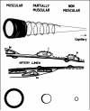
Schematic showing the graded structural changes in the media of the pulmonary arterial wall, moving from muscular arteries to precapillary non-muscular vessels. The top panel illustrates the gradual loss of musculature. The middle panel shows the transition from vascular smooth muscle cells (M) to intermediate cells (I) and finally to pericytes (P). The bottom panel represents cross-sections, highlighting the partial circumferential coverage of the media. From Reid et al. (252), with permission.
Figure 7
Figure 7
Organization of vascular smooth muscle in small partially muscular vessels elucidated with immunohistochemistry. In light micrographs of rat lung (5 μm sections, hematoxylin counterstain), vascular smooth muscle can be seen to partially cover the vascular wall. Smooth muscle cells staining positive for CD40, seen in cross-section (A), cover ~ 50% of the vessel wall perimeter. Similar coverage is seen in a more superficial section immunostained for vascular cell adhesion molecule or VCAM (B). In the latter image, some broad bands of smooth muscle cells are organized in a spiral (arrowhead), rather than circumferential, fashion over the wall surface. Scale 10 μm in both panels.
Figure 8
Figure 8
Endothelial ultrastructure in the arterial wall. Endothelial cells (A, scale 500 nm, rat lung) connected by a long tight junctional complex (TJ) are situated on a broad elastic lamina which separates the intima of this vessel from vascular smooth muscle cells (VSM) in the media. Numerous vesicles and caveolae, as well as mitochondria and Weibel-Palade bodies (*) are present. In an enlarged section from this image (B), microtubules (arrowheads) and a Weibel-Palade body (*) can be seen adjacent to the interendothelial cell tight junction. The very dense, laminated appearance of the junctional complex at the basal cell border denotes a gap junction (GJ). A small Golgi network (GN) sits adjacent to the nucleus (C, mouse lung).
Figure 9
Figure 9
Ultrastructure of vascular smooth muscle. Two micrographs from mouse lung show smooth muscle cells in the arterial wall with myofibrils along the long-axis of the cell (A, 500 nm scale) and when the cell is sectioned perpendicular to this plane (B, 500 nm scale). In the latter view, myofibrils appear as lighter clumps surrounded by mitochondria and rough endoplasmic reticulum. At the media-adventitia border (A), a fibroblast (F) containing an extensive rough endoplasmic reticulum network and the vascular smooth muscle cell (VSM) lie on either side of the external elastic lamina (EL). Bundles of fibrillar collagen (COL) are seen in the adventitia as well. Numerous dense bodies (A, arrowhead) are evident in the smooth muscle cell. A higher resolution image (C) shows the fine structure of an extensive parallel array of myofibrils (MF) in a smooth muscle cell. Panel D shows a gap junction (GJ) connecting two smooth muscle cells in mouse pulmonary artery.
Figure 10
Figure 10
Stellate fibroblasts in the vascular wall. The stellate nature and extensive cytoplasmic projections of adventitial fibroblasts are better appreciated in the adventitia of an extra-alveolar vessel from edematous mouse lung.
Figure 11
Figure 11
Freeze-fracture of inter-endothelial junctions. Freeze-fracture replicas of endothelium from a small intra-acinar pulmonary artery (A) and a pulmonary capillary (B). In the artery, packed clusters of gap junction particles (arrowheads) lie embedded between junctional strands. In contrast, the inter-endothelial junction in the capillary is much simpler, comprised of only 1-2 junctional strands. From Schneeberger and Lynch (268), with permission.
Figure 12
Figure 12
Myoendothelial junctions in the pulmonary vasculature. Various morphologies in myoendothelial junctions (arrowheads) can be found in rat (A and B) or mouse (C) lung. Either the endothelial cell or the smooth muscle cell (or both) sends a projection through a focal discontinuity in the internal elastic lamina, allowing contact. VSM, vascular smooth muscle; EL, elastic lamina; COL, collagen fibrils. Scales are 1 μm.
Figure 13
Figure 13
Variable origin of pulmonary capillary networks. Precapillary arteries may branch at right angles from a small parent artery then give rise to a capillary network after some distance (A and B). Alternatively small arteries may abruptly end in a capillary network (C and D). Capillary networks may also emerge directly from the parent artery (E). In human and rat lung, extra-alveolar pulmonary arteries 100 μm or more in diameter may give rise directly to capillary networks (119, 137). From Horsfield, with permission (119).
Figure 14
Figure 14
Capillary density differs in perivascular and pleural networks versus that in the alveolar septal wall. Vascular corrosion casting of rat lung has elucidated variability in capillary density in the distal lung. Low density networks are present adjacent to extra-alveolar vessels (A) and on the pleural surface (B); scale bars are 100 μm. In contrast, high density capillary networks populate alveolar septal walls (C, scale 50 μm). Panel A from Guntheroth et al. (98), with permission.
Figure 15
Figure 15
Endothelium in alveolar capillaries can be extremely attenuated. In this section from rat lung, the thin side of the septal wall is shown for a capillary running across a septal wall fold. No alveolar space can be seen. Both the endothelium (EC) and the type I alveolar epithelium (EPI) are extremely attenuated, with few organelles present other than vesicles and an occasional mitochondria. Through much of this section the basement membranes of these layers appear fused, except at the tip of the fold (upper right). While interstitium is quite thin, nonetheless several interstitial cells are apparent. A pericyte (P) lies adjacent to the endothelium and is enveloped by the endothelial basement membrane. Attenuated cytoplasmic processes of several interstitial fibroblasts are present, one containing a lipid droplet (L). Scale 1 μm.
Figure 16
Figure 16
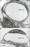
Capillary ultrastructure in rat and mouse lung. In cross section, the very attenuated endothelial cells comprising the capillary can be seen (A, scale 2 μm, from rat lung). In this section, portions of three cells can be identified. For the most part, the perinuclear region typically evident on the thick side of the septal wall remains out of the section plane for this capillary. While the presence of edema in the septal wall is not normal, this image does allow clear visualization of collagen fibers (COL) and interstitial cells, including fibroblasts (F). Long, thin cytoplasmic extensions of the fibroblast wrap around nearly half of the capillary circumference. In addition, a pericyte (P) can be seen closely adherent to the basal aspect of one endothelial cell. The attenuated type I alveolar epithelial cells (T1) and a type II epithelial cell (T2) complete the septal wall facing the alveolus (Alv). The capillary shown in panel B (scale 1 μm) is situated at the tip of a septal wall, where typically bundles of collagen (COL) and elastin (EL) fibers provide support at the entrance ring to the alveolus. In this view, thick and thin sides of the alveolar septal wall are more clearly delineated. A fibroblast (F) containing an extensive rough endoplasmic reticulum and lipid droplets (L) is present within the interstitium. In panels A and B, red dashed boxes that highlight inter-cellular connections in the septal wall are enlarged in Figure 17.
Figure 17
Figure 17
Intercellular connections in the alveolar septal wall. Boxed areas from Figure 16 are enlarged here. In panel A, the thin cytoplasmic extension of a interstitial fibroblast (F) approaches the alveolar type I epithelium (T1). Panel B shows tight junctions between type I and type II (T2) alveolar epithelial cells. Finally, panel C shows the close apposition between an interstitial fibroblast and the capillary endothelium (EC). Arrows show points of cell-cell contact. Similar interconnectedness has been documented in human lung (287); see Figure 18 for more detail.
Figure 18
Figure 18
Model of intercellular connections in the alveolar septal wall. This model of the alveolar septal wall was developed using serial sections of human lung, visualized with transmission electron microscopy. Type I (T1) and type II (T2) alveolar epithelial cells interdigitate at the lateral borders of the type II cell (sites 1 and 7). Intercellular connections between the type II cell and fibroblasts (F) in the alveolar septal wall take on numerous morphologies (sites 2-6), enabled by focal discontinuities in the type II cell basement membrane. Fibroblasts also connect with pericytes (P, site 8), type I cells (site 9), and capillary endothelial cells (En, site 10). From Sirianni et al. (287), with permission.
Figure 19
Figure 19
Thickness and composition of the alveolar septal wall interstitium. The total thickness of the alveolar septal wall in adult mammals scales with body size (and alveolar curvature, not shown), although the slope of the relationship is relatively shallow. In addition, the relative contributions of collagen and elastin in the septal wall interstitium also increase with body size and scale with alveolar curvature. “Other” includes cellular elements, basement membrane and other non-collagen/non-elastin areas within the interstitial compartment. Note that these data were calculated for the whole width of the alveolar septum, thus the measures of interstitial thickness shown here are 2-fold higher that measured as the arithmetic mean interstitial thickness (see Table 3). From Mercer et al., with permission (191).
Figure 20
Figure 20
Ultrastructure of pulmonary veins in rat and mouse. The wall of a large intrapulmonary vein from rat lung (Panel A, scale 3 μm) includes the endothelium (EC), scant smooth muscle (SM), fibroblasts (F), nerves (arrow) and abundant striated cardiac myocytes (CM). The pulmonary myocardium can be oriented in a circumferential or longitudinal pattern (or both as shown here). When multiple layers exist, they are much more loosely organized than are layers of smooth muscle cells in pulmonary arteries. Capillaries lie within the myocardial layer. In a pulmonary vein from mouse (panel B, scale 2 μm), an additional cell is present: the interstitial Cajal-like cell (ICLC).
Figure 21
Figure 21
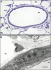
Resolution of the pulmonary venous myocardium. With light microscopy, a myocardial sleeve in intrapulmonary veins can appear similar to smooth muscle, unless smooth muscle-specific probes are utilized. Light (A, scale 25 μm) and electron (B, scale 2 μm) micrographs of a small pulmonary vein (lumen diameter ~ 100 μm) from the same block of rat lung highlight this point. For light microscopy, a 1 μm section of plastic embedded glutaraldehyde-fixed lung prepared for transmission electron microscopy was stained with toluidine blue. The media in this image is notable only by its relative thickness and density. A transmission electron micrograph of an 80 nm section from the same block elucidated the presence of a cardiac myocyte rather than smooth muscle in the media. PV, lumen of the pulmonary vein; Alv, alveolar space.
Figure 22
Figure 22
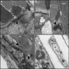
High resolution ultrastructure of pulmonary vein in rat and mouse lung. Panels A and B show myocytes in the venous myocardium cut through the short or long axis, respectively. Panel A shows desmosomes (arrowhead), seen as dense laminated junctional complexes structures, joining lateral borders of adjacent myocardial cells. In the long axis view (panel B), intercalated discs that connect myocytes end-to-end are apparent (arrowhead). The well-organized contractile apparatus, including clear I, Z and M bands, can be clearly seen in this image. Panel C shows dense segments of the plasmalemmal membrane (arrows) in a pulmonary vein smooth muscle cell adjoining a small tapered striated myocyte, which suggests the potential for gap junction communication. Finally, panel D shows a small nerve bundle (N) in the adventitia of pulmonary veins, adjacent to striated myocytes. The axons are enclosed in Schwann cells (S). EC, endothelial cell; VMS, vascular smooth muscle cell; CM, cardiac myocyte. Scale 1 μm.
Figure 23
Figure 23
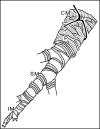
Model of pulmonary vein structure. Using a combination of light and scanning EM, Hashizume and colleagues developed a model of the pulmonary vein (106). After capillaries coalesce into small non-muscular pulmonary veins, several compartments appear sequentially in the venous tree. These include 1) partially muscular pulmonary veins populated by intermediate (IM) and vascular smooth muscle cells (SM) and 2) muscular veins, where a sleeve of striated cardiac myocytes (CM) may overlay the smooth muscle. This model has general utility for understanding structure in pulmonary veins, with the caveat that in large mammals, myocardial sleeves only appear in large extra-pulmonary veins (107, 117, 216, 219, 220, 320). In contrast, in rat and mouse, the musculature of even small muscular intrapulmonary veins is comprised of smooth muscle and a sleeve of striated myocardium (151, 170, 206, 216, 230). When present, the myocardium is overlaid (and intertwined) with a capillary network and nerve fibers. From Hashizume et al. (106), with permission.

References

    1. Adachi T, Ishikawa K, Hida W, Matsumoto H, Masuda T, Date F, Ogawa K, Takeda K, Furuyama K, Zhang Y, Kitamuro T, Ogawa H, Maruyama Y, Shibahara S. Hypoxemia and blunted hypoxic ventilatory responses in mice lacking heme oxygenase-2. Biochem Biophys Res Commun. 2004;320:514–522. 10.1016/j.bbrc.2004.05.195 S0006291X04012100 [pii] - PubMed
    1. Aguirre JI, Morrell NW, Long L, Clift P, Upton PD, Polak JM, Wilkins MR. Vascular remodeling and ET-1 expression in rat strains with different responses to chronic hypoxia. Am J Physiol Lung Cell Mol Physiol. 2000;278:L981–987. - PubMed
    1. Aharinejad S, Bock P, Lametschwandtner A, Firbas W. Scanning and transmission electron microscopy of venous sphincters in the rat lung. Anat Rec. 1992;233:555–568. 10.1002/ar.1092330410. - PubMed
    1. Aharinejad S, Egerbacher M, Nourani F, Bock P, Friederici C, Schraufnagel DE. Pulmonary venous sphincters in cattle. Anat Rec. 1996;246:356–363. 10.1002/(SICI)1097-0185(199611)246:3<356::AID-AR6>3.0.CO;2-W [pii] 10.1002/(SICI)1097-0185(199611)246:3<356::AID-AR6>3.0.CO;2-W. - PubMed
    1. Aiello VD, Gutierrez PS, Chaves MJ, Lopes AA, Higuchi ML, Ramires JA. Morphology of the internal elastic lamina in arteries from pulmonary hypertensive patients: a confocal laser microscopy study. Mod Pathol. 2003;16:411–416. 10.1097/01.MP.0000067685.57858.D7. - PubMed

Publication types

MeSH terms

LinkOut - more resources