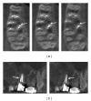
Artefacts in Cone Beam CT Mimicking an Extrapalatal Canal of Root-Filled Maxillary Molar
- PMID: 23606995
- PMCID: PMC3626249
- DOI: 10.1155/2013/797286
Artefacts in Cone Beam CT Mimicking an Extrapalatal Canal of Root-Filled Maxillary Molar
Abstract
Despite the advantages of cone-beam computed tomography (CBCT), the images provided by this diagnostic tool can produce artifacts and compromise accurate diagnostic assessment. This paper describes an endodontic treatment of a maxillary molar where CBCT images suggested the presence of a nonexistent third root canal in the palatal root. An endodontic treatment was performed in a first maxillary molar with palatal canals, and the tooth was restored with a cast metal crown. The patient returned four years later presenting with a discomfort in chewing, which was reduced after occlusal adjustment. CBCT was prescribed to verify additional diagnostic information. Axial scans on coronal, middle, and apical palatal root sections showed images similar to a third root canal. However, sagittal scans demonstrated that these images were artifacts caused by root canal fillings. A careful interpretation of CBCT images in root-filled teeth must be done to avoid mistakes in treatment.
Figures


References
-
- Vertucci FJ. Root canal morphology and its relationship to endodontic procedures. Endodontic Topics. 2005;10(1):3–29.
-
- Torabinejad M, Walton R. Endodontics: Principles and Practiceedition. 4th edition. St. Louis, Mo, USA: Saunders/Elsevier; 2009.
-
- Vertucci FJ. Root canal anatomy of the human permanent teeth. Oral Surgery, Oral Medicine, Oral Pathology, Oral Radiology and Endodontology. 1984;58(5):589–599. - PubMed
-
- de Deus QD, Horizonte B. Frequency, location, and direction of the lateral, secondary, and accessory canals. Journal of Endodontics. 1975;1(11):361–366. - PubMed
-
- Gulabivala K, Aung TH, Alavi A, Ng YL. Root and canal morphology of Burmese mandibular molars. International Endodontic Journal. 2001;34(5):359–370. - PubMed
LinkOut - more resources
Full Text Sources
Other Literature Sources
Miscellaneous

