

Case Reports
doi: 10.4103/2156-7514.107995.
Print 2013.
Mixed adenoneuroendocrine carcinoma of cecum: a rare entity
Affiliations
- PMID: 23607079
- PMCID: PMC3625879
- DOI: 10.4103/2156-7514.107995
Item in Clipboard
Case Reports
Mixed adenoneuroendocrine carcinoma of cecum: a rare entity
J Clin Imaging Sci.
.
Abstract
Mixed adenoneuroendocrine carcinoma of cecum (MANEC) was first reported by Cardier in 1924. These tumors are thought to arise from multi-potential stem cells, which have differentiated bidirectionally. Location of the tumor influences the treatment and outcome. We report a rare case of MANEC where the patient presented with abdominal pain and distension. Imaging revealed an ileo colic intussusception with the lead point being a MANEC.
Keywords: Cecum; composite tumor; mixed adenoneuroendocrine carcinoma.
Conflict of interest statement
Figures

Aditi Jain
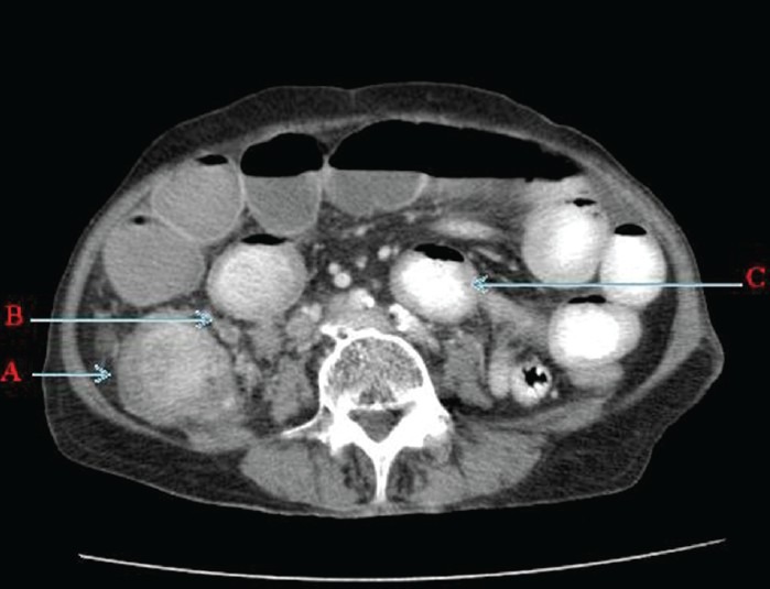
Contrast enhanced computed tomography of abdomen and pelvis (axial section) shows A. Enhancing Intraluminal caecal mass with caecal wall thickening. B. Adjacent necrotic mesentric nodes and C. Dilated small bowel loops with few of them having thickened walls.
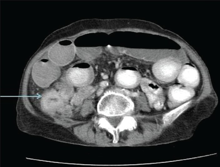
Contrast enhanced computed tomography of abdomen and pelvis (axial section) reveals thickened ileum loop extending into the cecum (arrow).
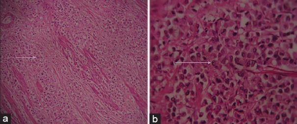
Hematoxylin and eosin stained sample (a) at ×10 and (b) at ×40 demonstrate large monomorphic tumor cells having large round cell nucleus and abundant eosinophilic cytoplasm (seen arranged in cords and nests in Figure 3a).
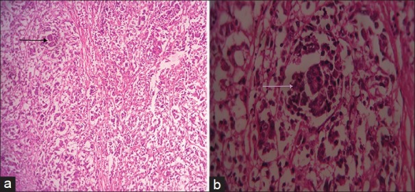
Hematoxylin and eosin stained tissue (a) at ×10 and (b) at ×40 show glandular formation (arrow) amounting to >30% of the tumor.
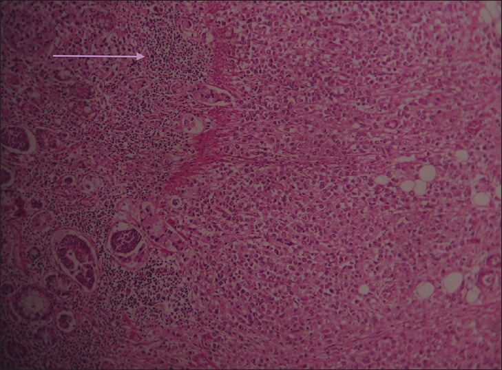
Hematoxylin and eosin stained tissue at ×10 shows tumor cells surrounded by focal lymphocyte response.
References
-
- Klimstra DS, Modlin IR, Coppola D, Lloyd RV, Suster S. The pathologic classification of neuroendocrine tumors: A review of nomenclature, grading, and staging systems. Pancreas. 2010;39:707–12. - PubMed
-
- Suttmann H. A mixed adenocarcinoma and neuroendocrine carcinoma of the rectum associated with an adenocarcinoma of the prostate as a second primary malignancy: Report of a case. Electron J Pathol Histol. 2003;9:32–8.
-
- Bosman FT, Carneiro F, Hruban RH, Theise ND. WHO Classification of Tumours of the Digestive System. 4th ed. Lyon, France: WHO, International Agency for Research on Cancer; 2010. Nomenclature and classification of neuroendocrine neoplasms of the digestive system; p. 13.
Publication types
LinkOut - more resources
Full Text Sources
Other Literature Sources
