. 2013 Aug;173(2):163-72.
doi: 10.1111/cei.12107.

Investigation of the cutaneous response to recall antigen in humans in vivo

Affiliations

Investigation of the cutaneous response to recall antigen in humans in vivo

A N Akbar et al. Clin Exp Immunol. 2013 Aug.

Abstract

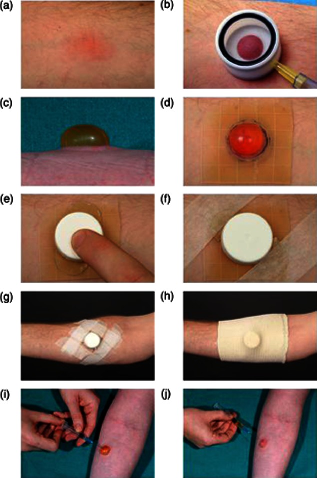
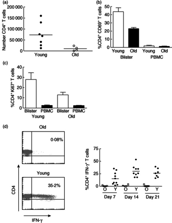
In this paper we provide a detailed description of an experimental method for investigating the induction and resolution of recall immune response to antigen in humans in vivo. This involves the injection of tuberculin purified protein derivative (PPD) into the skin, followed by inducing suction blisters at the site of injection, from which leucocytes and cytokines that are involved in the response can be isolated and characterized. Using this technique we found that although the majority of CD4(+) T cells in the skin that are present early in the response express cutaneous lymphocyte antigen (CLA), the expression of this marker is reduced significantly in later phases. This may enable these cells to leave the skin during immune resolution. Furthermore, interleukin (IL)-2 production can be detected both in CD4(+) T cells and also in the blister fluid at the peak of the response at day 7, indicating that mediators found in the blister fluid are representative of the cytokine microenvironment in vivo. Finally, we found that older humans have defective ability to respond to cutaneous PPD challenge, but this does not reflect a global immune deficit as they have similar numbers of circulating functional PPD-specific CD4(+) T cells as young subjects. The use of the blister technology enables further characterization of the skin specific defect in older humans and also general mechanisms that govern immune regulation in vivo.

Keywords: DTH; ageing; human immunity.


Figures

Fig. 1
Fig. 1
Practical aspects of skin suction blister induction. (a) Skin suction blisters were raised on Mantoux tests or normal skin 18–24 h prior to sampling. (b) A suction cup was centred on the Mantoux test and a negative pressure of 25–40 kPa (200–300 mmHg) below atmospheric pressure was applied to the skin until (c) a unilocular blister was formed. The blister was dressed with (d) a 5 × 5-cm piece of Comfeel Plus ulcer dressing, (e) a trimmed universal container top, (f,g) Micropore tape and (h) a Tubigrip bandage. (i–j) Blister fluid was aspirated the following day using a sterile 23-G needle and 2-ml syringe.
Fig. 2
Fig. 2
Blister volumes and leucocyte numbers isolated from skin suction blisters raised over Mantoux tests (MT) 7 days after induction. Figures show data collected from 30 skin suction blisters raised on day 7 following MT induction. (a) Blister fluid volume aspirated compared to MT induration diameter (mm) at day 7. (b) Number of leucocytes isolated from skin suction blisters at day 7 compared to the clinical response measured at day 3. (c) Number of leucocytes isolated per microlitre (μl) of skin suction blister fluid at day 7 compared to the clinical response measured at day 3. The Spearman's rank correlation (r) and P-values are shown for each graph.
Fig. 3
Fig. 3
Characteristics of skin-infiltrating T cells following Mantoux test (MT) induction. (a) Cutaneous lymphocyte antigen (CLA) expression on peripheral blood and normal skin blister CD3+ T cells. Peripheral blood mononuclear cells (PBMC) and blister cells were isolated and stained with anti-CD3, anti-CD4- and anti-CLA antibodies and analysed by flow cytometry. Samples were analysed by gating on the CD3+CD4+ CD45RA subset of cells within the live lymphocyte/lymphoblast gate. Gating strategy was used to exclude cell debris, but include lymphocyte blasts. Dot-plots showing CLA expression on CD3+ T cells isolated from the peripheral blood (PB) and a suction blister raised on day 3 following MT induction (b) PBMC (◊) and suction blister (SB) (▪) cells isolated from skin following MT induction were analysed using flow cytometry for the percentage of CD4+ memory T cells positive for CLA expression. The mean ± standard error of the mean of three experiments per time-point is shown.
Fig. 4
Fig. 4
Antigen-specific cells accumulating following Mantoux test (MT) induction make interleukin (IL)-2 and interferon (IFN)-γ. (a) Blister cells were stimulated with purified protein derivative (PPD) ex vivo for 15 h in the presence of brefeldin A. CD4+ T cells were then examined for intracellular IFN-γ and IL-2 expression by flow cytometry. Representative fluorescence activated cell sorter (FACS) plots are shown on the left. The percentage of CD4+ T cells expressing IFN-γ (black bars), IL-2 (white bars) and both IFN-γ and IL-2 (grey bars) are shown. The mean ± standard error of the mean (s.e.m.) of three to four experiments per time-point is shown. (b) IL-2 expression in suction blister (SB) supernatants was assayed by multiplex bead immunoassay using a Luminex 100 and cytokine Beadlyte assay kit. The graph shows the mean ± s.e.m. of three to 18 experiments per time-point. The dotted line on the graph showing IL-2 data denotes the lower limit of detection in the assay.
Fig. 5
Fig. 5
Old individuals do not mount a good clinical response to Mantoux test despite showing equivalent responses in circulating CD4 T cells. (a) Comparison of clinical scores between young and old. Twenty-five young (under 40) and 32 old (aged more than 70 years) individuals had a Mantoux test. Induration diameter, erythema index and palpability of the lesion were assessed and graded at day 3 and a clinical score assigned based on these parameters. (b) Comparison of the proportion of PPD-specific cells in peripheral blood of old and young individuals. PBMCs were collected from 35 young and 40 old individuals and stimulated with PPD ex vivo for 15 h in the presence of brefeldin A. CD4+ T cells were then examined for intracellular interferon (IFN)-γ. Representative fluorescence activated cell sorter (FACS) plots are shown on the left. The percentage of CD4+ T cells expressing IFN-γ in young and old individuals is shown in the graph (n = 18 young, 31 old). (c) PBMC from young and old individuals were stimulated ex vivo with PPD (1 μg/ml) for 6 days. Proliferation was assessed by [3H]-thymidine incorporation.
Fig. 6
Fig. 6
Reduced clinical responses to Mantoux test (MT) in old individuals are associated with reduced T cell accumulation and activation in the skin. (a) Skin suction blisters were raised on day 7 following MT induction in young and old individuals. Absolute number of CD4+ T cells in each blister was calculated using fluorescence activated cell sorter (FACS) analysis using Truecount tubes (n = 7 young and n = 4 old). (b) Cells isolated from skin suction blisters and peripheral blood on day 7 after the MT induction were stained with antibodies for CD3, CD4 and CD69 immediately ex vivo and analysed by flow cytometry. Graph shows the mean ± standard error of the mean of the percentage of CD4+ T cells expressing CD69 in blisters and peripheral blood mononuclear cells (PBMC) (Mann–Whitney U-test P = 0·0357; n = 4 young, 5 old). (c) Blister cells and PBMCs isolated on day 7 following MT were stained with antibodies for CD4 and Ki67 in order to identify CD4+Ki67+ T lymphocytes by flow cytometry (Mann–Whitney U-test P = 0·095; n = 5, mean ± standard error of the mean is shown). (d) Blister cells were collected at different times following MT induction and stimulated with purified protein derivative (PPD) ex vivo for 15 h in the presence of brefeldin. CD4+ T cells were then examined for intracellular interferon (IFN)-γ expression by flow cytometry. Representative FACS plots are shown on the left. The percentage of CD4+ T cells producing IFN-γ at different time-points is shown in the graph (horizontal lines represent the mean).

References

    1. Davis MM. A prescription for human immunology. Immunity. 2008;29:835–838. - PMC - PubMed
    1. Hayday AC, Peakman M. The habitual, diverse and surmountable obstacles to human immunology research. Nat Immunol. 2008;9:575–580. - PubMed
    1. Vukmanovic-Stejic M, Reed JR, Lacy KE, Rustin MH, Akbar AN. Mantoux test as a model for a secondary immune response in humans. Immunol Lett. 2006;107:93–101. - PubMed
    1. Vukmanovic-Stejic M, Agius E, Booth N, et al. The kinetics of CD4Foxp3 T cell accumulation during a human cutaneous antigen-specific memory response in vivo. J Clin Invest. 2008;118:3639–3650. - PMC - PubMed
    1. Reed JR, Vukmanovic-Stejic M, Fletcher JM, et al. Telomere erosion in memory T cells induced by telomerase inhibition at the site of antigenic challenge in vivo. J Exp Med. 2004;199:1433–1443. - PMC - PubMed

Publication types

MeSH terms